
Home ![]() MAYFAIR DIAGNOSTICS MAYFAIR PLACE RE-INTRODUCES ECHOCARDIOGRAMS
MAYFAIR DIAGNOSTICS MAYFAIR PLACE RE-INTRODUCES ECHOCARDIOGRAMS
Mayfair Diagnostics is pleased to announce the re-introduction of echocardiography imaging services at our Mayfair Place location, effective June 19, 2023.
We are again offering this service, interpreted by our fellowship-trained radiologists, in response to the demand from the community and to more comprehensively serve our patients. Mayfair’s cardiac imaging services now include echocardiography, exercise stress testing, and myocardial perfusion imaging (MPI), as well as private pay cardiac-specific CT coronary calcium scoring and CT coronary angiography. All cardiac services are offered at our Mayfair Place location.
Mayfair Diagnostics Mayfair Place offers all medical imaging modalities: bone density, breast imaging, cardiac imaging, computed tomography (CT), magnetic resonance imaging (MRI), nuclear medicine imaging, pain management, ultrasound, and X-ray services.
Located in unit 132 at 6707 Elbow Drive SW, Mayfair Place is located on the corner of Glenmore Trail and Elbow Drive SW, at the south end of the Mayfair Place apartment building.
Our Mayfair Place clinic is open from 7 a.m. to 9 p.m., Monday to Thursday, 7 a.m. to 4 p.m. on Fridays, and 8 a.m. to 4 p.m. on Saturdays. You will need an imaging requisition from your health care practitioner for all exams. Walk-in X-ray patients don’t require an appointment. Other exams can be booked at the clinic, through our online appointment request option, or by calling our central booking number at 403-777-3000.
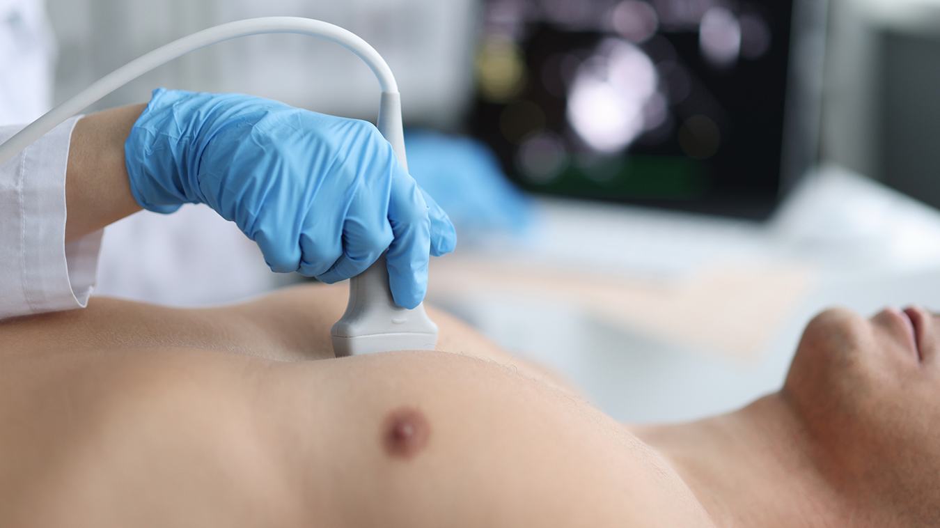
For symptoms like shortness of breath, heart palpitations, or chest pain, your doctor might request a heart ultrasound to help find the cause for your concerns. Also known as an echocardiogram or ECHO, this type of medical imaging uses a wand-like transducer, or probe, that emits high-frequency sound waves to create an image of your heart.
Your image will show the shape and movement of your heart valves, as well as the size of your heart chambers and how well they are working. It can help investigate your clinical symptoms and assess heart conditions, such as murmurs or damage to the heart due to prior heart attack or infection.
A heart ultrasound is a non-invasive way to get a look at how well the heart is working. It can be used on its own or in conjunction with an exercise stress test. During an exercise stress test, you are connected to a monitor and your blood pressure and heart rate are observed as you exercise on a treadmill. By monitoring and recording changes in heart rate and blood pressure, this test can indicate if parts of the heart have inadequate blood supply and evaluate how well your heart performs under stress, which is an important indicator of heart health.
This exam may also be ordered along with myocardial perfusion imaging (MPI), or a nuclear stress test, in which a radioactive material (radiopharmaceutical) is injected intravenously and you are monitored to see how well it is taken up by your heart muscle as it flows through the heart arteries at rest and during exercise.
An echocardiogram is most often ordered in cases with symptoms, especially if you have a family history of heart disease or other high risk factors for heart disease. If you have a family history of heart disease, it’s important to talk with your health care practitioner about your heart health, including how to monitor it or address any other risk factors that can be changed.
During an echocardiogram your chest area will need to be bare, so you will be asked to change into a gown and lie down on an examination bed. A registered cardiac sonographer will apply small electrocardiograph patches and a scentless, hypoallergenic ultrasound gel to your chest.
Your sonographer will move the transducer across your chest and a computer will convert echoes from sound waves into pictures of your heart onto a screen. You will be asked to change positions from your back to your side to view the heart from different angles, and you may experience some pressure from the transducer in order to optimize the images.
Throughout the exam, you will hear noises that may sound like your heartbeat, although it’s not your heart beating you will hear. This Doppler sound is assessing the movement of blood through the heart chambers and valves.
After your exam is complete, your results will be interpreted by a radiologist and your health care practitioner will receive a comprehensive report within a few days of your exam. Your doctor will then be able to review your results with you, along with the results of any other test you may have undergone, and determine the next steps in your health care treatment plan.
To determine whether this exam is appropriate for you, you will need to discuss your symptoms and medical history with your health care practitioner, who would then provide you with a requisition for this procedure if it’s indicated. You can then call to book an appointment with our Contact Centre and they will explain how to prepare for your exam.
Please visit our cardiac imaging page for more information about what happens during this exam and how to prepare.
REFERENCES
Heart and Stroke Foundation (2023) “Echocardiogram.” www.heartandstroke.ca. Accessed May 24, 2023.
Mayo Clinic Staff (2023) “Echocardiogram.” www.mayoclinic.org. Accessed May 25, 2023.
© 2024 Mayfair. All rights reserved.
| Cookie | Duration | Description |
|---|---|---|
| cookielawinfo-checkbox-analytics | 11 months | This cookie is set by GDPR Cookie Consent plugin. The cookie is used to store the user consent for the cookies in the category "Analytics". |
| cookielawinfo-checkbox-functional | 11 months | The cookie is set by GDPR cookie consent to record the user consent for the cookies in the category "Functional". |
| cookielawinfo-checkbox-necessary | 11 months | This cookie is set by GDPR Cookie Consent plugin. The cookies is used to store the user consent for the cookies in the category "Necessary". |
| cookielawinfo-checkbox-others | 11 months | This cookie is set by GDPR Cookie Consent plugin. The cookie is used to store the user consent for the cookies in the category "Other. |
| cookielawinfo-checkbox-performance | 11 months | This cookie is set by GDPR Cookie Consent plugin. The cookie is used to store the user consent for the cookies in the category "Performance". |
| viewed_cookie_policy | 11 months | The cookie is set by the GDPR Cookie Consent plugin and is used to store whether or not user has consented to the use of cookies. It does not store any personal data. |