
Mammography is still the gold standard for breast cancer screening. However, there are other breast imaging services that are beneficial when used in conjunction with mammography, such as digital breast tomosynthesis and automated breast ultrasound.
For example, digital breast tomosynthesis (DBT) is a type of 3D mammography that uses computerized detection plates that take multiple images in an arc. This provides pictures from numerous different angles. Digital technology combines the images, adding them together and sharpening certain areas and blurring others. This produces a more detailed image that helps to better illuminate the presence of cancers, without increased radiation levels or scanning time.
All Mayfair locations use DBT. Plus, in 2018, Mayfair Diagnostics was the first in Western Canada to install the leading-edge Senographe Pristina Dueta mammography system in several of our clinics. Now we have 13 mammography clinics with this new system, which uses DBT.
Pristina also helps make mammograms more comfortable for patients through its ergonomic design and a special feature called patient-assisted compression. After the breast is properly positioned by a technologist and initial compression is set, you can choose to use a handheld wireless remote control to adjust the level of compression to what’s comfortable for you, under the guidance of a technologist.
Some patients find mammograms uncomfortable, due to the pressure, and are therefore less likely to go for regular breast cancer screening with mammography. Patient-assisted compression allows the patient to reduce the pressure of the exam, while still maintaining excellent diagnostic quality. Giving patients some measure of control during their exam can help decrease their anxiety and increase their willingness to return.
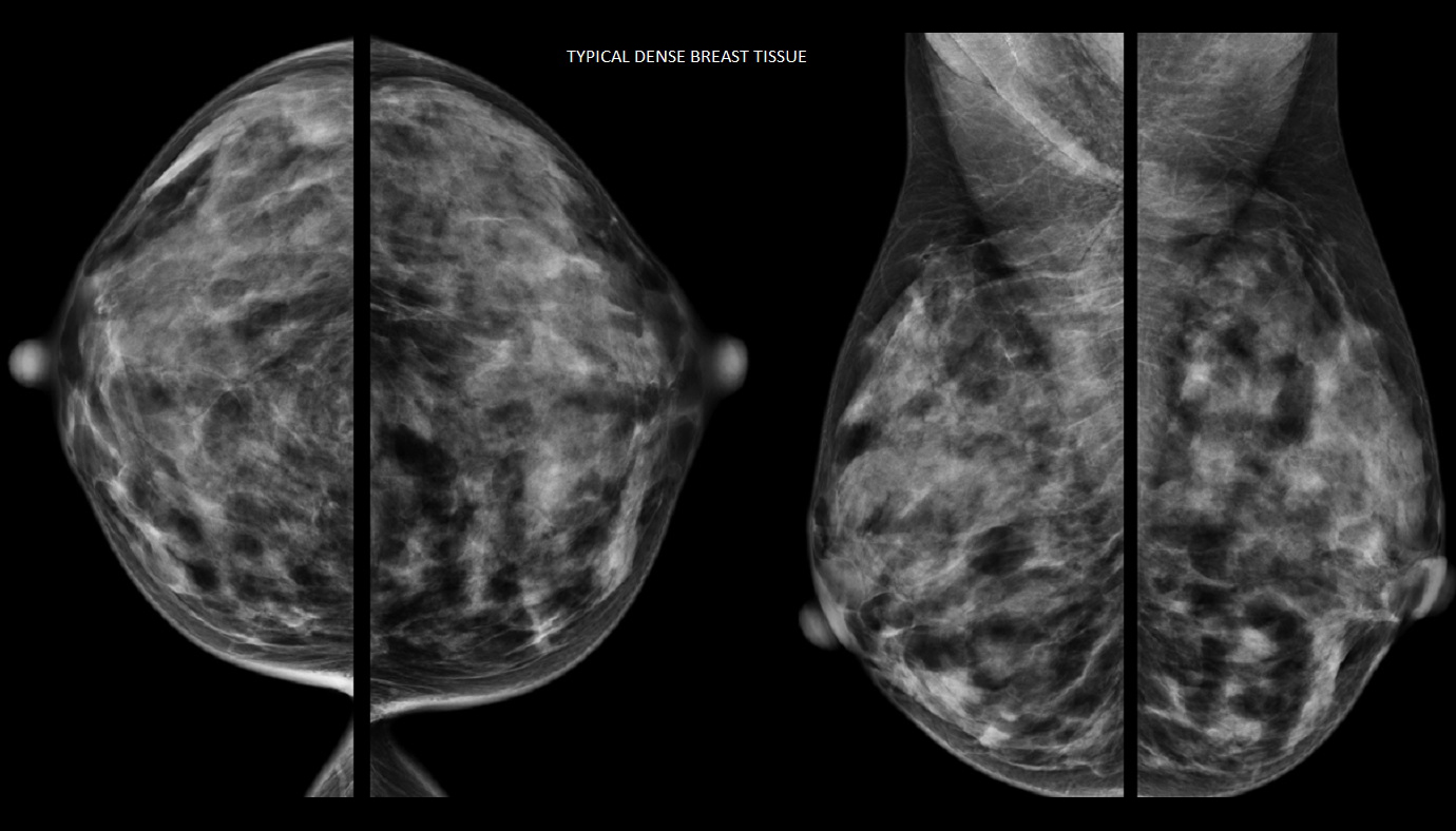
This higher level of detail is especially important when scanning dense breasts, because the more dense tissue that is present the higher the risk for breast cancer. Breast density is not a measure of how breasts look or feel, rather it describes how well an x-ray travels through the tissue.
On a mammogram, fatty tissue looks dark, while both dense tissue and tumours look white, making it hard to differentiate between the two. The white-looking breast cancers are easier to see on a mammogram when they’re surrounded by dark-looking fatty tissue.
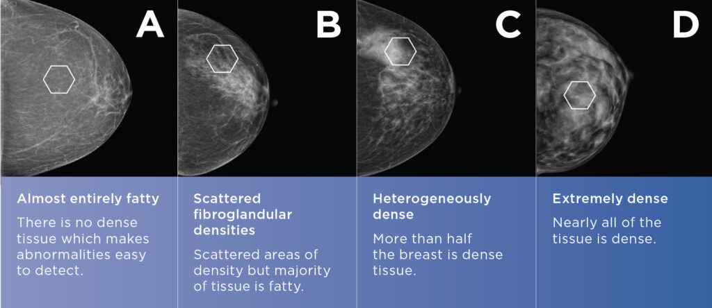
The Breast Imaging Reporting and Database Systems (BI-RADS), classifies breast density into four groups, or breast density scores:
Scoring is not an exact science and radiologists often disagree about levels of density. Mayfair uses the Volpara Health Technologies program which scores density from A to D.
Automated breast ultrasound (ABUS) is another breast imaging service that is especially beneficial to women with dense breast tissue. A high breast density (Volpara) score can make a mammogram harder to read and ABUS allows the technologist to check the breast from a variety of angles offering more accurate interpretations. For extremely dense (Volpara D), or heterogeneously dense breast tissue (Volpara C) combined with additional risk factors for breast malignancy, Mayfair recommends supplemental ABUS or handheld breast ultrasound.
With ABUS, images are acquired with a large transducer that is placed on the breast in several different positions. They are then processed at a workstation and reviewed, removing operator bias and providing a uniform assessment process. ABUS is also fast and almost painless, looking at both breasts in about 15 minutes.
A number of studies indicate that ultrasound is one of the most promising adjunct screening modalities because of its benefit for dense breasts and its low cost and availability.
Mayfair offers DBT at all 14 of our mammography locations and patient-assisted compression at 13 of our clinics. Coventry Hills has a mammography system that uses DBT but doesn’t offer patient-assisted compression.
Automated breast ultrasound is available at our Market Mall, Mayfair Place, Southcentre, and The CORE locations. Please visit our breast imaging services page for more information.