Several studies have looked at the benefits of adding handheld breast ultrasound and automated breast ultrasound (ABUS) to a screening mammogram. Results have shown that it increases cancer detection and sensitivity, particularly in cases of women with dense breasts.
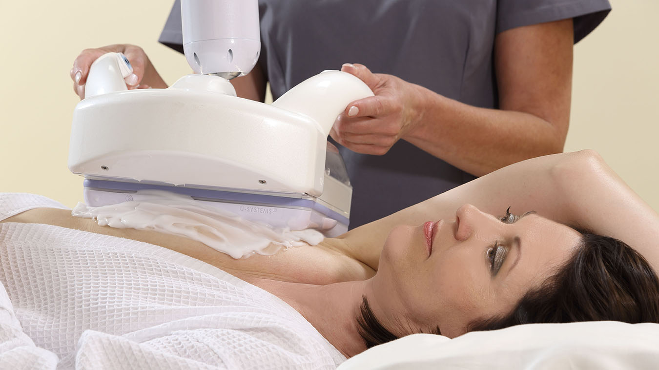
Automated breast ultrasound uses high frequency sound waves to take 3D images of the whole breast, targeting specific areas and ensuring they are displayed in a reproducible fashion. It’s a fast and almost painless procedure which can look at both breasts in about 15 minutes. ABUS is especially beneficial to women with dense breast tissue.
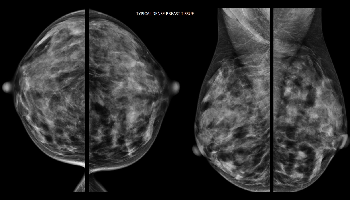
You can’t tell by look or feel whether or not you have dense breasts. It’s a diagnosis that can only be assessed by mammography. Dense breasts have less fat and more glandular and connective tissue. On a mammogram, fatty tissue looks dark, while both dense tissue and tumours look white, making them hard to detect. The white-looking breast cancers are easier to see on a mammogram when they’re surrounded by dark-looking fatty tissue.
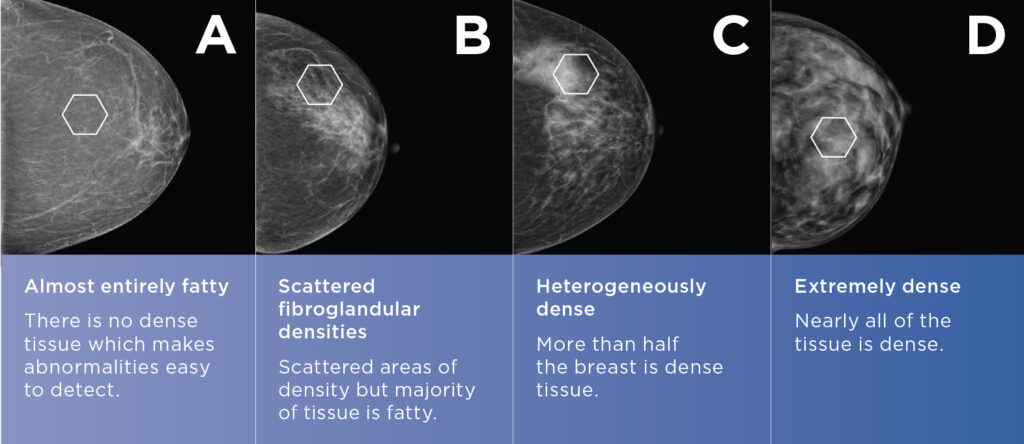
For women with dense breast, a mammogram may be harder to read, so smaller cancers may be hidden. At Mayfair Diagnostics, we use the Volpara Health Technologies program which scores breast density from A to D:
In cases of high breast density, ABUS allows the technologist to check the breast from a variety of angles offering more accurate interpretations. For extremely dense (Volpara D), or heterogeneously dense breast tissue (Volpara C) combined with additional risk factors for breast malignancy, Mayfair recommends supplemental ABUS or handheld breast ultrasound, in addition to regular mammograms.
Dense breast tissue increases the risk of breast cancer, especially in older women. For women with dense breast tissue in 75 percent or more of their breasts, their breast cancer risk is double relative to the general population.
Mammography is still the gold standard for breast cancer screening. However, women with very dense breasts may require a more personalized screening approach than what is recommended for the general population. This may include both mammography and ultrasound exams.
Mayfair Diagnostics has 14 mammography locations, which have all been updated recently with the newest technology. Thirteen of our mammography locations also feature patient-assisted compression – which helps provide a more comfortable mammogram. Coventry Hills has an upgraded mammography system but doesn’t offer patient-assisted compression.
Automated breast ultrasound is available at our Market Mall, Mayfair Place, Southcentre, and The CORE locations. Please visit our breast imaging services page for more information.
Advani, S. M., et al. (2021) “Association of Breast Density With Breast Cancer Risk Among Women Aged 65 Years or Older by Age Group and Body Mass Index.” JAMA Network Open. 2021;4(8). Accessed October 20, 2022.
Alberta Health Services Breast Cancer Screening Programs (2022) “Breast Cancer Screening.” www.screeningforlife.ca. Accessed October 20, 2022.
Canadian Cancer Society (2022) “Breast cancer statistics.” www.cancer.ca. Accessed October 20, 2022.
Canadian Cancer Society (2022) “Breast density.” www.cancer.ca. Accessed October 20, 2022.
Eske, J. (2019) “How do the breasts change with age and why?” www.medicalnewstoday.com. Accessed October 20, 2022.
Łuczyńska, E., et al. (2022) “The Role of ABUS in The Diagnosis of Breast Cancer.” Journal of Ultrasonography. 2022 Apr; 22(89): 76–85. Accessed October 20, 2022.