Home HOW DOES A CHEST X-RAY HELP DIAGNOSE BREATHING PROBLEMS?
If you were asked what you would see in an X-ray image, odds are you would say bones. And it’s true – X-ray imaging is very good at looking at your bones.
But interestingly, the most frequently performed X-ray exam is a chest X-ray. This exam may be ordered to look for rib fractures or other problems with bones in the chest, but it’s also frequently requested to look at the lungs and heart.
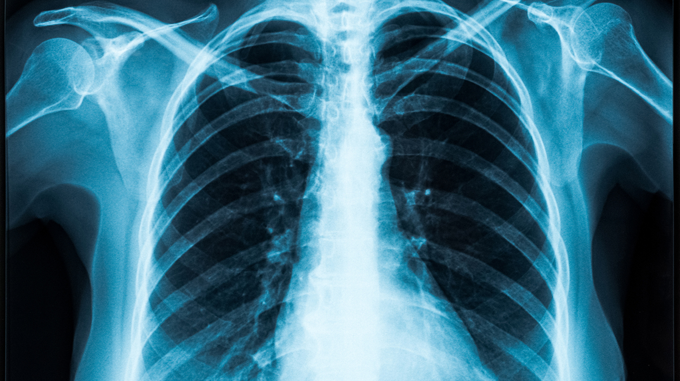
X-ray imaging uses a type of electromagnetic ionizing radiation to create images of your lungs, heart, and the bones of your chest and spine. A small amount of radiation is sent into the chest and different areas of the chest absorb this radiation at different rates, creating an image with various shades of grey. When filled with air, the lungs show up as darker areas while the heart and lung vessels will appear as lighter areas.
Different lung and heart pathologies can be determined by the radiologist depending on their appearance within the lungs. For example, infections in the lungs can show up as white patchy areas. A chest X-ray can help diagnose such lung concerns as:
Your doctor might order a chest X-ray for a number of reasons, including:
Mayfair Diagnostics performs about 40,000 chest X-rays each year. During this exam, you will be asked to change into a gown and to remove any metal objects, such as jewelry and clothes with metal buttons, zippers, or snaps. We may also ask you to tie up any long hair that may obstruct a clear view of the chest.
If you are a woman between the ages of 11-55, you will be asked about the possibility of pregnancy prior to your exam, since X-rays use radiation. Lead shielding can also be provided to cover vulnerable areas.
One of our experienced technologists will help you move into different positions standing against an upright board. You will be asked to turn to the front and side and move your arms and shoulders into various positions. Our state-of-the-art imaging technology will take views of the front and side of your chest. You will also be required to take a deep breath and hold it for several seconds to help your heart and lungs show up more clearly. We need the lungs filled with air in order to assess them.
The X-ray itself is not painful but holding a particular position may be uncomfortable if you are experiencing pain. Overall, the exam may take about 15 minutes, although once in the X-ray room the process typically takes less than five minutes.
Once your doctor has identified the need for an exam, you will be given a requisition form. X-rays are offered in the community on a walk-in basis at any medical imaging clinic, including 12 Mayfair Diagnostics locations in Calgary and one in Regina. Appointments are not required for general X-ray procedures; simply bring your form with you.
Your X-ray images will be reviewed by a specialized radiologist who will compile a report that is sent to your doctor within 1-2 business days, sooner for urgent requests. Mayfair Diagnostics is owned and operated by over 50 radiologists who are fellowship-trained in many key areas, such as neuroradiology, body, cardiac, musculoskeletal imaging, etc. This allows for an expert review of your imaging by the applicably trained radiologist.
Your images will also be uploaded to a provincial picture archiving and communication system (PACS) – this technology provides electronic storage and convenient access to your medical images from multiple sources, such as your doctor, specialists, hospitals, walk-in clinics, etc.
Please note, X-ray services are NOT offered at our Cochrane, Saskatoon, Southcentre, and Sunpark locations. Visit our locations page to view clinics near you.
DerSarkissian, C. (2020) “Is It Bronchitis or Pneumonia?” www.webmd.com. Accessed July 13, 2021.
Healthwise Staff (2020) “Chest X-Ray.” www.myhealth.alberta.ca. Accessed July 13, 2021.
Mayo Clinic Staff (2021) “Chest X-rays.” www.mayoclinic.org. Accessed July 13, 2021.
Nabili, S. N. (2020) “What Does a Chest X-Ray Show?” www.medicinenet.com. Accessed July 13, 2021.
We foster a supportive and collaborative culture designed to encourage positive patient experiences and build strong working relationships across the organization:
Our core values shape the way we work with patients, partners, and fellow employees. And, more than anything else, they’re what set Mayfair apart. In everything we do, this is what we strive for:
EXCELLENCE
We share a commitment to high quality and excellence in all that we do. This commitment calls on all of us to achieve the very best of our capabilities and exceed our own expectations.
CURIOSITY
We innovate in everything, from services to processes. We believe meaningful change and effective problem solving come only by looking at challenges and opportunities from new angles and by exercising our creativity and curiosity.
PASSION
We show pride, enthusiasm, and dedication in everything that we do. We are committed to producing and delivering high-quality results and services. We are passionate about our industry and about our company, services, partners, and patients.
COLLABORATION
Our team is supportive of each other’s efforts; we are loyal to one another; and we care for one another both personally and professionally. We promote and support a diverse, yet unified, team. We work together to meet our common goals across Mayfair clinics, locations, and geographies. Only through collaboration on ideas, technologies, and talents can we achieve our mission and vision.
SERVICE
We take pride in delivering exceptional service every day. We listen to every request with an open mind, always looking for opportunities to go above and beyond to create memorable, personalized experiences. We take responsibility to answer our referrers’ and patients’ requests and respect their time by always responding with a sense of urgency.
Start a career with Mayfair Diagnostics — one of Western Canada’s leading medical imaging teams.
Headquartered in Calgary, Alberta, we’ve been helping people f ind clarity for their health for over 100 years. At our clinics in Calgary and area, Regina, and Saskatoon, our team of radiologists, technologists, and support staff work in a truly integrated way to provide exceptional experiences for our patients. Joining our team is more than a job. It’s an investment in your future — a plan for success.
OUR PEOPLE
Our people share our quest to make a difference in our patient’s lives. We’re a team of professionals, disciplined in our skills and compassionate with our patients, providing the care and attention they need. At our core, we are a trusted partner in our patients’ health care journey. Our patients, physicians, and other health care providers rely on us for quality imaging to help manage their patient’s health care decisions with certainty. But our business is about more than just imaging. It’s about building lasting relationships and making a meaningful difference in the lives of those we meet.
OUR VISION
A world in which every person has clarity about their health. We push the boundaries of what is possible and embrace change as an opportunity. We strive to be thought leaders and encourage creativity by providing a safe place for calculated risk taking. We learn from our mistakes. We share best practices across our operations and are recognized by our peers for our work. We engage the best to help propel us forward in achieving our goals.