TOLL FREE:
1-866-611-2665

Home HOW DOES ULTRASOUND WORK?
Ultrasound imaging uses high-frequency sound waves to create an image of the inside of your body. It’s very good a looking at the soft tissues of the body and is often the first step in determining the cause for your symptoms.
Also known as sonography, ultrasound imaging uses a small transducer (probe) to both transmit sound waves into the body and record the waves that echo back. Sound waves travel into the area being examined until they hit a boundary between tissues, such as between fluid and soft tissue, or soft tissue and bone. At these boundaries some of the sound waves are reflected back to the probe, while others travel further until they reach another boundary and are reflected back. Since the speed, direction, and distance sound waves travel differ depending on the boundary they run into, a computer can interpret this information as a two-dimensional image on a screen.
The shape and intensity of the echoes depend on how the area absorbs the sound waves. For example, most waves pass through a fluid-filled cyst and send back very few or faint echoes, which look black on the display screen. On the other hand, waves will bounce off a solid tumor, creating a pattern of echoes that the computer will interpret as a lighter-colored image. Air and bone also reflect sound waves.
Ultrasound has been around for over sixty years and is considered safe since there are no known risks and it doesn’t use radiation. It’s one of the most commonly ordered imaging exams since it’s versatile, portable, relatively inexpensive, non-invasive, and can provide real-time information about the area of concern.
Ultrasound has a variety of uses, despite being most often associated with pregnancy. It can be ordered to investigate pain, swelling, or other symptoms.
For example, ultrasound can help determine the composition of a lump, distinguishing between a cyst and a tumour. A cyst is a sac filled with fluid, which is mostly benign. A tumour is an area of complex tissue, which can be either benign or malignant. Ultrasound can usually help differentiate between benign and malignant tumours based on shape, location, and a number of other sonographic characteristics. Both cysts and tumours can be found in your skin, tissue, organs, and bones.
Ultrasound is a standard part of prenatal care, providing images of the fetus or information on the embryo’s viability and growth.
An abdominal ultrasound can help check for kidney stones, gallstones, liver disease, and the cause of stomach pain. Multiple still images are taken to represent the location, texture, and blood flow of each organ.
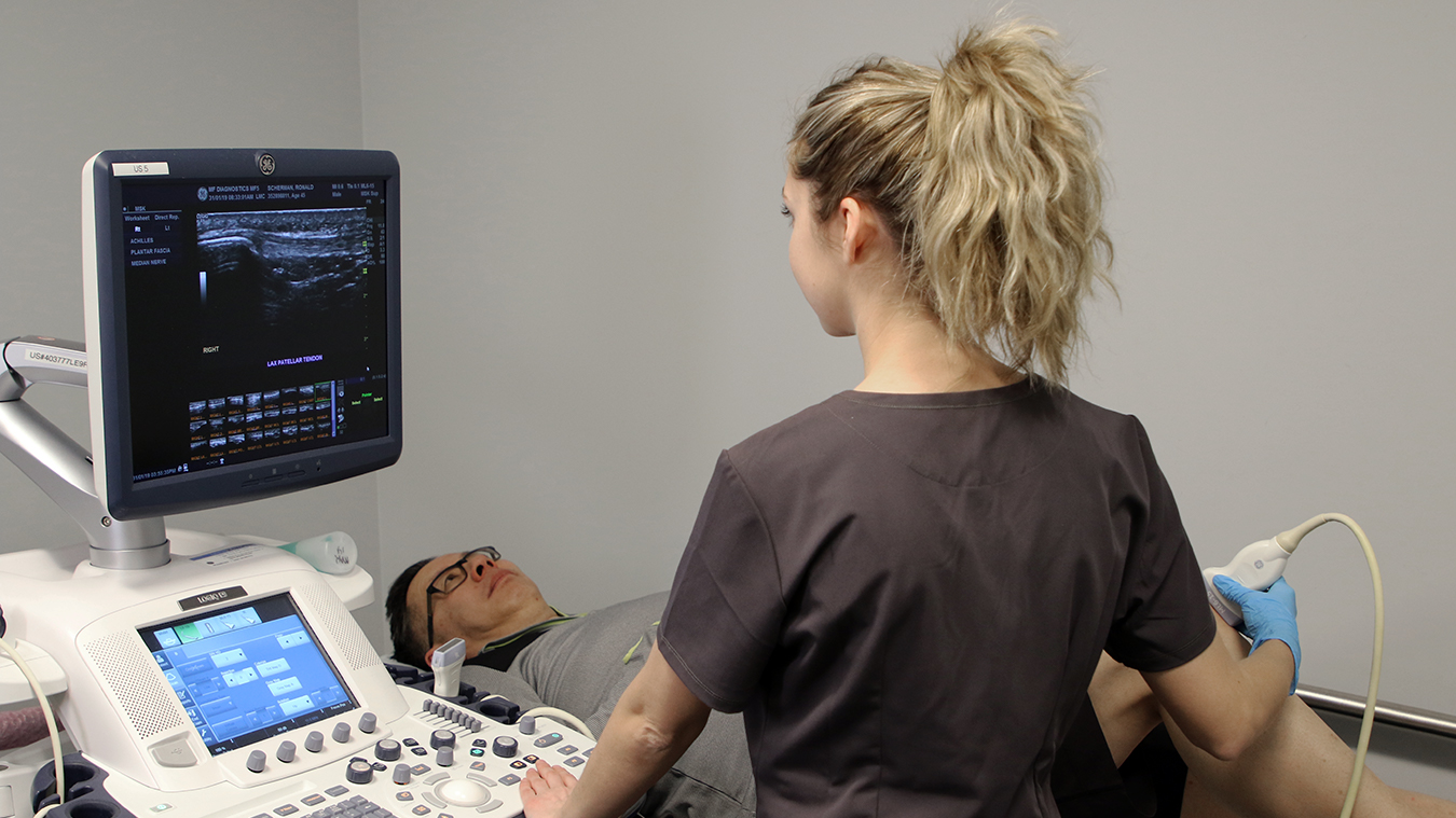
Ultrasound is also very good at looking at cartilage, muscles, tendons, and ligaments to evaluate joints for fluid or inflammation. Called a musculoskeletal (MSK) ultrasound, these exams are often ordered for joint concerns, such as symptoms in the ankle, elbow, knee, shoulder, or wrist. For these exams the dynamic nature of ultrasound is an advantage for accurate diagnosis, since we can evaluate the area in question while it’s moving and watch as a patient performs the action causing symptoms. MSK ultrasounds may be requested on their own or in conjunction with an X-ray to rule out a fracture.
Ultrasound imaging is covered under the Alberta and Saskatchewan Health Care Insurance Plans. It helps health care practitioners make a diagnosis and inform care decisions. Once your doctor has identified the need for an ultrasound, your doctor’s office may book an appointment for you, or provide you with a number to call to book your appointment.
Some ultrasound exams require preparation before the exam. You will be provided with preparation instructions before your exam, or you can visit our website for more information about your specific exam.
For example, for an abdominal ultrasound, you will be asked to fast and have nothing to eat or drink (except water) for six hours prior to your exam. For some obstetrical ultrasounds, you will need to arrive with a full bladder.
Your images will be reviewed by a specialized radiologist who will compile a report that is sent to your doctor within 24 hours, sooner for urgent requests. Mayfair Diagnostics is owned and operated by over 50 radiologists who are fellowship-trained in many key areas, such as neuroradiology, body, cardiac, musculoskeletal, etc. This allows for an expert review of your imaging by the applicably trained radiologist.
Your images will be uploaded to a provincial picture archiving and communication system (PACS) – this technology provides electronic storage and convenient access to your medical images from multiple sources, such as your doctor, specialists, hospitals, and walk-in clinics.
Your doctor will review your images and the report from the radiologist and discuss next steps with you, such as a treatment plan or the need for further diagnostic imaging or lab tests to ensure an accurate diagnosis.
Mayfair Diagnostics has 13 locations across Calgary which provide ultrasound services, as well as one in Cochrane and one in Regina. For more information, please visit our services page or call our toll-free number 1-866-611-2665.
REFERENCES
American Cancer Society (2015) “Ultrasound for Cancer.” www.cancer.org. Accessed March 3, 2020.
Brazier, Y. (2017) “How do ultrasound scans work?” www.medicalnewstoday.com. Accessed March 3, 2020.
Freudenrich, C.C. (2001) “How ultrasound works.” University of Toronto Physics. Accessed March 3, 2020.
Lewis, T. (2013) “5 Fascinating Facts About Fetal Ultrasounds.” Live Science. www.livescience.com. Accessed March 3, 2020.
Mayo Clinic Staff (2022) “Abdominal Ultrasound.” www.mayoclinic.org. Accessed March 3, 2020.
O’Keefe Osborn, C. (2018) “What’s the Difference Between Cysts and Tumors?” www.healthline.com. Accessed March 3, 2020.
Radiological Society of North America (2020) “General Ultrasound.” www.radiologyinfo.org. Accessed March 3, 2020.
Our Refresh newsletter delivers the latest medical news, expert insights, and practical tips straight to your inbox, empowering you with knowledge to enhance patient care and stay informed.
By subscribing to our newsletter you understand and accept that we may share your information with vendors or other third parties who perform services on our behalf. The personal information collected may be stored, processed, and transferred to a country or region outside of Quebec.
Please read our privacy policy for more details.