Home ![]() WHEN IS BREAST MRI APPROPRIATE?
WHEN IS BREAST MRI APPROPRIATE?
More women are surviving a breast cancer diagnosis thanks to early detection and more effective treatment options. In 2017, 89% of women diagnosed with breast cancer were still alive after five years.
While it’s easy to understand the importance of breast screening when looking at survival rates, understanding the different breast screening technologies and when to use them can be confusing. Mammography, breast ultrasound, and breast magnetic resonance imaging (MRI) can all be used to help diagnose breast cancer, or other breast diseases.
A mammography exam takes X-ray images – mammograms – of the breast to look for abnormalities. It provides a detailed view of breast tissue with a very small dose of radiation and can reveal changes that may be too small for you or your doctor to feel. It is the gold standard for breast screening and is usually the first step in the screening process.
Breast ultrasound uses sound waves to check breast tissue from a different perspective than mammography. It can be handheld or an automated breast ultrasound, which uses 3D ultrasound technology to offer a fast and reproducible look at the breast from a variety of angles. Breast ultrasounds are often requested when you or your health care practitioner feel a lump, see nipple discharge, or your mammogram shows new findings. Ultrasound can also be ordered when your mammogram shows high breast density. They are often performed as a supplement to a mammography exam.
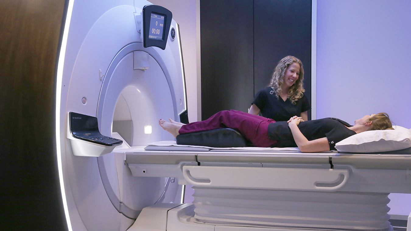
Breast MRI uses a powerful magnetic field and radio waves to take a very detailed look at the soft tissues of the breast. Although mammography is still the standard first method of detecting abnormalities in breast tissue, Breast MRI can be a powerful screening and diagnostic tool for women at high risk of breast cancer. It can also be used to assess the extent of breast tumors after a cancer diagnosis, for further examination of concerns when mammography and/or ultrasound are negative, or as a follow up after treatment to look for recurrence of breast cancer. Women with extremely dense breast tissue may also benefit from breast MRI.
Your risk for breast cancer depends on a number of factors including your personal medical history, age, genetics, lifestyle, etc. Discussing your risk factors with your health care practitioner will help you gauge your risk level.
The following are considered high risk factors:
NOTE: If you are at high risk, Mayfair recommends regular breast screening every year starting at any age above 40 or 10 years earlier than the age a first-degree relative was diagnosed with breast or ovarian cancer.
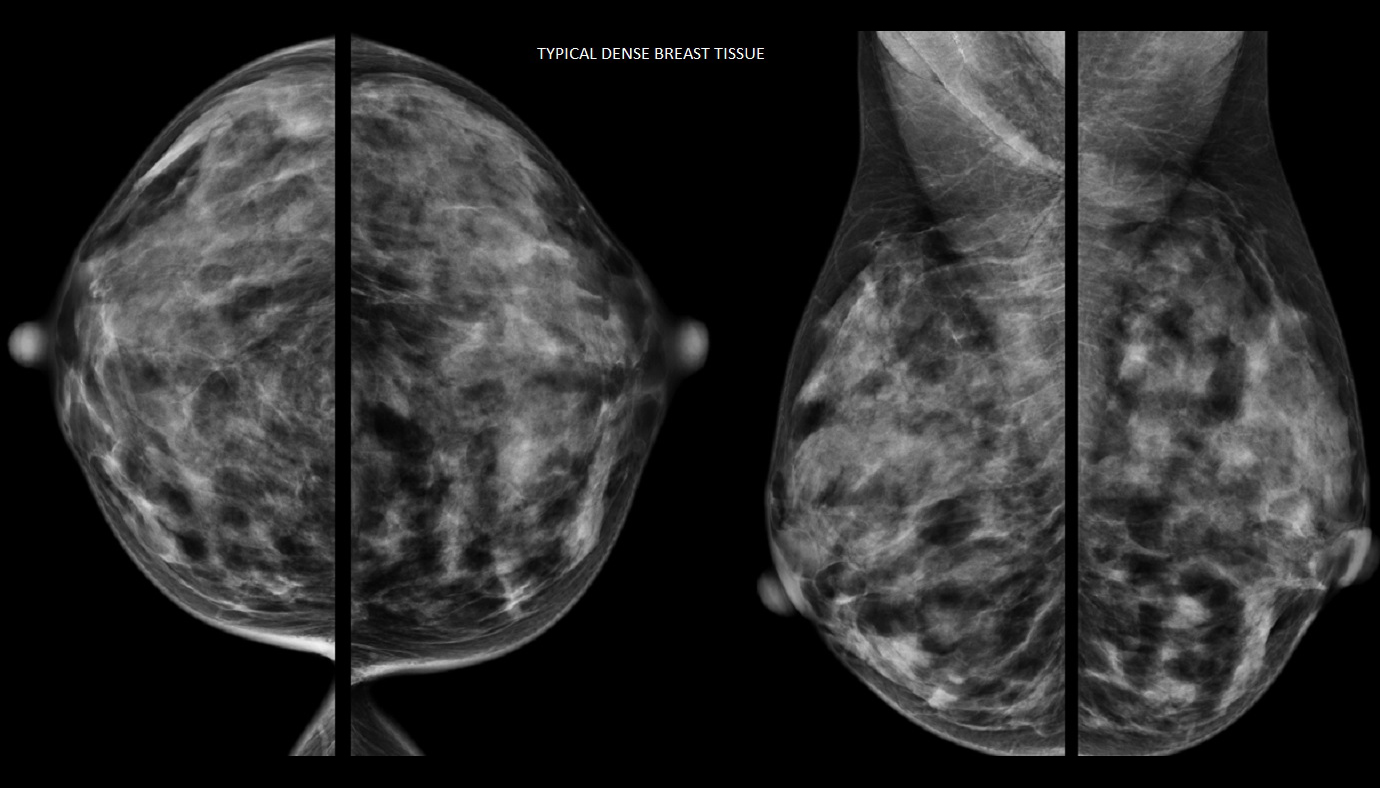
*Breast density is a major risk factor for breast cancer. Your breasts are made up of different types of tissue: fibroglandular (dense) tissue, and fat (not dense tissue). Dense breasts have less fatty tissue, more fibroglandular tissue, and a higher risk of cancer. Fatty tissue appears dark on a mammogram while both abnormalities and fibroglandular tissue appear white, making abnormalities harder to find.
Dense breasts are quite common, but they can only be determined by a mammogram. At Mayfair Diagnostics all of our mammography machines are equipped with software that classifies breast density, which is included in reports to referring doctors. It’s important to know your breast density and discuss it with your doctor since women with dense breast tissue often benefit from regular mammograms supplemented by ultrasound or possibly breast MRI.
How mammography, breast ultrasound, and breast MRI work together and whether they would all be appropriate depends on your personal circumstances and will need to be determined by your doctor.
Mayfair Diagnostics has 13 mammography locations with patient-assisted compression – which helps provide a more comfortable mammogram. Coventry Hills has an upgraded mammography system but doesn’t offer patient-assisted compression.
Automated breast ultrasound is available at our Market Mall, Mayfair Place, Southcentre, and The CORE locations. Please visit our breast imaging services page for more information.
REFERENCES
Advani, S. M., et al. (2021) “Association of Breast Density With Breast Cancer Risk Among Women Aged 65 Years or Older by Age Group and Body Mass Index.” JAMA Network Open. 2021;4(8). Accessed October 8, 2022.
Alberta Health Services Breast Cancer Screening Programs (2022) “Breast Cancer Screening.” www.screeningforlife.ca. Accessed October 8, 2022.
Canadian Cancer Society (2021) “Breast cancer statistics.” www.cancer.ca. Accessed October 8, 2022.
Canadian Cancer Society (2021) “Breast density.” www.cancer.ca. Accessed October 8, 2022.
Engmann et al. (2017) “Population-Attributable Risk Proportion of Clinical Risk Factors for Breast Cancer.” JAMA Oncology. 2017; 3(9) 1228-36. Accessed October 8, 2022.
Huizen, J. (2019) “What happens during a breast MRI?” www.medicalnewstoday.com. Accessed on October 8, 2022.
Tabar, L., et al. (2018) “The incidence of fatal breast cancer measures the increased effectiveness of therapy in women participating in mammography screening.” Cancer. Accessed on October 8, 2022.
We foster a supportive and collaborative culture designed to encourage positive patient experiences and build strong working relationships across the organization:
Our core values shape the way we work with patients, partners, and fellow employees. And, more than anything else, they’re what set Mayfair apart. In everything we do, this is what we strive for:
EXCELLENCE
We share a commitment to high quality and excellence in all that we do. This commitment calls on all of us to achieve the very best of our capabilities and exceed our own expectations.
CURIOSITY
We innovate in everything, from services to processes. We believe meaningful change and effective problem solving come only by looking at challenges and opportunities from new angles and by exercising our creativity and curiosity.
PASSION
We show pride, enthusiasm, and dedication in everything that we do. We are committed to producing and delivering high-quality results and services. We are passionate about our industry and about our company, services, partners, and patients.
COLLABORATION
Our team is supportive of each other’s efforts; we are loyal to one another; and we care for one another both personally and professionally. We promote and support a diverse, yet unified, team. We work together to meet our common goals across Mayfair clinics, locations, and geographies. Only through collaboration on ideas, technologies, and talents can we achieve our mission and vision.
SERVICE
We take pride in delivering exceptional service every day. We listen to every request with an open mind, always looking for opportunities to go above and beyond to create memorable, personalized experiences. We take responsibility to answer our referrers’ and patients’ requests and respect their time by always responding with a sense of urgency.
Join Mayfair Diagnostics, recognized as one of Western Canada’s premier medical imaging organizations. With a century-long legacy and headquartered in Calgary, Alberta, Mayfair Diagnostics is dedicated to assisting patients in achieving clarity regarding their health.
Operating clinics in Calgary and surrounding areas, Regina, and Saskatoon, our multidisciplinary team of radiologists, technologists, and support staff collaborate seamlessly to deliver high quality patient care. Working here is more than just a job; it’s a strong step towards your future.
OUR TEAMS
We are a dedicated group of professionals who combine skill with compassion to deliver attentive care to our patients. As a reliable partner in their health care journey, we provide high-quality imaging that assists patients, physicians, and other providers in making informed health decisions. Our work goes beyond imaging; it’s about fostering relationships and positively impacting everyone we serve.
OUR VISION
We envision a world where every person has clarity about their health. We innovate and welcome change, promoting leadership and creativity through safe risk-taking. We share best practices, earn peer recognition for our work, and engage top talent to reach our goals.
We strive to be thought leaders and encourage creativity by providing a safe place for calculated risk taking.
We share best practices across our operations and are recognized by our peers for our work. We engage the best to help propel us forward in achieving our goals.