
Home ![]() WHY WOULD I NEED A SCROTUM ULTRASOUND?
WHY WOULD I NEED A SCROTUM ULTRASOUND?
The scrotum is a pouch of skin containing the testicles, nerves, and blood vessels. Your health care practitioner might request a scrotum ultrasound to investigate an abnormality within the scrotum or the cause for symptoms like lumps, swelling, or pain. It could also be used to help diagnose trauma to the scrotal area, evaluate the cause of infertility, or look for an undescended testicle.
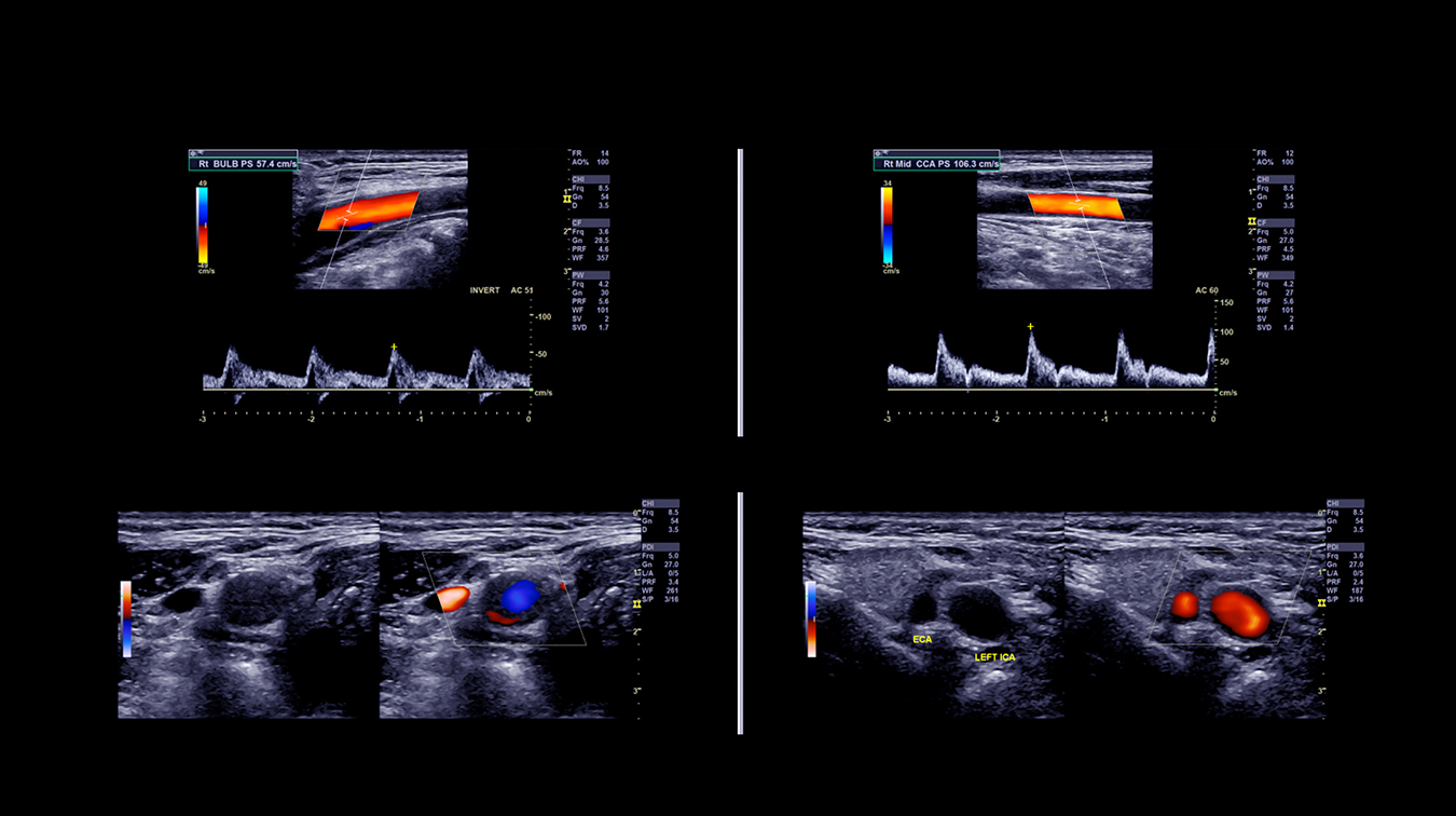
Ultrasound imaging uses high-frequency, real-time sound waves to create an image. A small transducer (probe) both transmits sound waves into the body and records the waves that echo back. The shape and intensity of the echoes depend on whether the area absorbs or transmits the sound waves. For example, most waves pass through a fluid-filled cyst and send back very few or faint echoes, which look black on the display screen. On the other hand, waves will bounce off a solid tumor, creating a pattern of echoes that the computer will interpret as a lighter-colored image. Air and bone also reflect sound waves.
Most scrotal lesions are benign. Ultrasound can usually help differentiate between benign and malignant lesions. If ultrasound identifies a lesion suspicious for malignancy, the findings are immediately conveyed to the referring physician. In these cases, the recommendation is for referral to the Urological Surgical Service for further management, which is usually surgical biopsy
Testicular cancer starts as an abnormal growth or tumour that develops in the cells of one or both of the testicles. These cells can change and no longer grow or behave normally, leading to benign or cancerous tumours. There are several types of testicular cancer, but the most common is a germ cell tumour.
Testicular cancer is the most common cancer in young Canadian men aged 15-35, but when caught and treated early the average survival rate is 97%, according to the Canadian Cancer Society. The incidence of testicular cancer diagnoses has been slowly increasing since 1984, but the mortality rate has been steadily decreasing. An estimated 1,200 Canadian men will be diagnosed with testicular cancer in 2022. An estimated 35 will die from the disease.
Because it’s one of the more survivable cancers, it’s important to be aware of your risk factors:
It’s also important to find out what’s normal for you and be aware of any changes. Find a warm environment and roll your testicles between your thumb and fingers to check for lumps or swelling. They should be smooth, firm, and comfortable to touch. It’s normal if one testicle is slightly bigger than the other, or if one hangs lower than the other. However, if your testicles feel heavier than usual, have changed in shape or size, or are painful, you should speak to your doctor.
Most lumps and bumps on your testicles are not cancer, but to investigate your doctor may book a scrotum ultrasound appointment for you, or provide you with a number to call to book your appointment. You will also be given a requisition form. There is no preparation required for your exam.
The exam usually takes 15-20 minutes. You will be asked to wear comfortable clothes. Once you arrive for your exam, you will be asked to remove your pants and underwear and lie down on the exam. A sheet will be draped across your lower body and the sonographer will apply a warm, non-scented, hypo-allergenic ultrasound gel. The scrotal area needs to be visible to the sonographer for correct orientation of the images.
You may experience mild to moderate pressure while the sonographer scans the right and left side of the scrotum. You may also be asked to indicate the area of concern by pointing out the lump or area of pain if it is localized to one spot. This will ensure that the correct area is properly assessed.
Once your exam is completed, your images will be reviewed by a specialized radiologist who will compile a report that is sent to your doctor within 24 hours, sooner for urgent requests. Mayfair Diagnostics is owned and operated by over 50 specially trained radiologists which allows for an expert review of your imaging by the applicably trained radiologist.
Your doctor will review your images and the report from the radiologist and discuss next steps with you, such as a treatment plan or the need for further diagnostic imaging or lab tests to ensure an accurate diagnosis.
Mayfair Diagnostics has 13 locations across Calgary, one in Cochrane and one in Regina, which offer ultrasound services. For more information about our clinic services, please visit our services page.
REFERENCES
Canadian Cancer Society (2022) “What is testicular cancer?” www.cancer.ca. Accessed November 5, 2022.
Canadian Cancer Society (2021) “Risk for testicular cancer.” www.cancer.ca. Accessed November 5, 2022.
Radiological Society of North America (2022) “Ultrasound – Scrotum.” www.radiologyinfo.org. Accessed October 10, 2019.
Radiologists are specialized physicians who interpret diagnostic imaging to diagnose and monitor a wide range of medical conditions. At Mayfair Diagnostics, they review X-ray, ultrasound, CT, and MRI studies, among others, analyzinsg images in detail and providing comprehensive reports and clinical recommendations to referring physicians. They collaborate closely with technologists and clinic teams to guide imaging protocols, ensure quality and radiation safety standards, and may perform image-guided procedures such as biopsies or injections. Through their expertise and teamwork, radiologists play a key role in accurate diagnosis, effective treatment planning, and improved patient outcomes.
Administrative professionals support the organization across human resources, marketing, operations, strategic partnerships, finance, information technology, and infrastructure. From recruiting staff to promoting services and improving workflows, they help ensure smooth operations and positive experiences for employees and patients. Through collaboration with clinical and support teams, they provide essential coordination that enables efficient, high-quality service.
Diagnostic Imaging Assistant (DIA) support clinic operations and help ensure a positive patient experience. They assist staff by greeting and preparing patients, confirming information, coordinating appointments, and guiding patients through their visit. DIAs also maintain exam rooms, manage documentation, and ensure supplies and equipment are ready. Through strong customer service, attention to detail, and teamwork, they help create a safe and organized environment.
Patient Experience Coordinators (PECs) are the first point of contact, scheduling exams and ensuring accurate patient information. They communicate clearly with patients, coordinate with care teams, and support a smooth, confidential, and customer-focused experience.
Nuclear Medical Technologists perform diagnostic nuclear medicine procedures involving sensitive and highly personal patient circumstances. They are responsible for delivering the highest standard of care in a professional, compassionate, and patient-centered manner, in accordance with provincial regulatory requirements, CAMRT standards, and Mayfair policies and guidelines.
Computed Tomography (CT) Technologists operate CT imaging equipment to produce detailed cross-sectional images of the body that assist in diagnosing a wide range of medical conditions. They prepare and position patients for scans, ensure safety protocols are followed, and administer contrast agents when required. CT technologists work closely with radiologists to ensure high-quality diagnostic images are obtained, while providing clear communication and compassionate care to support patient comfort throughout the procedure.
Magnetic Resonance Imaging (MRI) Technologists operates MRI scanners to produce detailed images of internal body structures used to assist in medical diagnosis and treatment planning. They are responsible for preparing and positioning patients, ensuring all safety protocols are strictly followed due to the strong magnetic field, and obtaining high-quality images as directed by radiologists. MRI technologists combine technical expertise with patient care, providing clear communication and support to ensure a safe, comfortable, and efficient imaging experience.
Diagnostic Medical Sonographers perform ultrasound exams to help diagnose and monitor medical conditions. Following Sonography Canada standards and Mayfair Diagnostics protocols, they capture accurate images while ensuring patient safety, comfort, and confidentiality. They work with radiologists and clinical teams to review requisitions, prepare patients, perform scans, and document findings, contributing to accurate diagnoses and a positive patient experience.
Medical Radiation Technologists (MRTs) perform x-ray, mammography, and BMD exams while ensuring patient safety, accuracy, and compassionate care. They also may assist in pain therapy procedures. MRTs verify patient information, explain procedures, position patients, and produce high-quality images. MRTs follow professional standards and protocols, maintaining strict radiation safety, quality assurance, and patient privacy while supporting a positive patient experience.
We foster a supportive and collaborative culture designed to encourage positive patient experiences and build strong working relationships across the organization:
Our core values shape the way we work with patients, partners, and fellow employees. And, more than anything else, they’re what set Mayfair apart. In everything we do, this is what we strive for:
EXCELLENCE
We share a commitment to high quality and excellence in all that we do. This commitment calls on all of us to achieve the very best of our capabilities and exceed our own expectations.
CURIOSITY
We innovate in everything, from services to processes. We believe meaningful change and effective problem solving come only by looking at challenges and opportunities from new angles and by exercising our creativity and curiosity.
PASSION
We show pride, enthusiasm, and dedication in everything that we do. We are committed to producing and delivering high-quality results and services. We are passionate about our industry and about our company, services, partners, and patients.
COLLABORATION
Our team is supportive of each other’s efforts; we are loyal to one another; and we care for one another both personally and professionally. We promote and support a diverse, yet unified, team. We work together to meet our common goals across Mayfair clinics, locations, and geographies. Only through collaboration on ideas, technologies, and talents can we achieve our mission and vision.
SERVICE
We take pride in delivering exceptional service every day. We listen to every request with an open mind, always looking for opportunities to go above and beyond to create memorable, personalized experiences. We take responsibility to answer our referrers’ and patients’ requests and respect their time by always responding with a sense of urgency.
Join Mayfair Diagnostics, recognized as one of Western Canada’s premier medical imaging organizations. With a century-long legacy and headquartered in Calgary, Alberta, Mayfair Diagnostics is dedicated to assisting patients in achieving clarity regarding their health.
Operating clinics in Calgary and surrounding areas, Regina, and Saskatoon, our multidisciplinary team of radiologists, technologists, and support staff collaborate seamlessly to deliver high quality patient care. Working here is more than just a job; it’s a strong step towards your future.
OUR TEAMS
We are a dedicated group of professionals who combine skill with compassion to deliver attentive care to our patients. As a reliable partner in their health care journey, we provide high-quality imaging that assists patients, physicians, and other providers in making informed health decisions. Our work goes beyond imaging; it’s about fostering relationships and positively impacting everyone we serve.
OUR VISION
We envision a world where every person has clarity about their health. We innovate and welcome change, promoting leadership and creativity through safe risk-taking. We share best practices, earn peer recognition for our work, and engage top talent to reach our goals.
We strive to be thought leaders and encourage creativity by providing a safe place for calculated risk taking.
We share best practices across our operations and are recognized by our peers for our work. We engage the best to help propel us forward in achieving our goals.