
Home ![]() WHEN WOULD I NEED A TRANSVAGINAL ULTRASOUND?
WHEN WOULD I NEED A TRANSVAGINAL ULTRASOUND?
For symptoms of pelvic pain, abdominal pain, unusual vaginal bleeding, difficulty conceiving, or other menstrual problems such as heavy or irregular periods, it’s important to speak with your health care practitioner. Your doctor may order a variety of tests to investigate the cause for these types of symptoms, including medical imaging such as a pelvic ultrasound with transvaginal ultrasound.
An ultrasound is a safe, versatile, and fast diagnostic imaging exam that uses high frequency sound waves transmitted through a transducer (probe) to produce images of your organs, tissues, and blood vessels.
A pelvic ultrasound is performed with a full bladder. During the exam sonographer will apply warm, non-scented ultrasound gel to your skin on your lower abdominal area before using a transducer to visualize the organs and surrounding area in your pelvis.
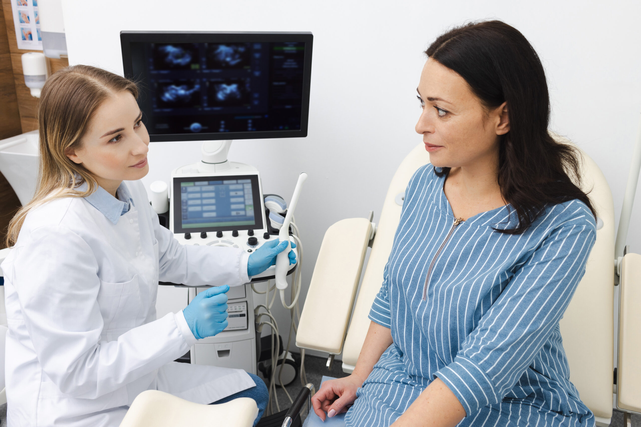
Whereas a transvaginal ultrasound (also known as an endovaginal ultrasound) uses a long, narrow transducer that looks like a wand is inserted into the vagina. This allows for a more detailed visual of the organs and the soft tissue inside the pelvic cavity.
A transvaginal ultrasound takes between 6-15 minutes. If you are having your period, the ultrasound can still be done. Prior to the exam, you will be asked to empty your bladder and will be given privacy to remove your clothes from the waist down and cover yourself with a large sheet. The sonographer will explain the procedure in detail and answer any questions to make sure you understand and consent to the exam.
For the exam the transducer is covered in a plastic sheath with lubricating gel that will be gently inserted into the vagina. Depending on your preference, you may be given the option to insert the transducer yourself, or the sonographer may do it for you. The sonographer will move the transducer around to capture photos. You may experience mild to moderate pressure while the sonographer takes the images, but the exam should not be painful. If you experience any pain or would like to discontinue the exam at any time, please let the sonographer know.
Your doctor may order this imaging to:
The technologist will not be able to provide any details or give any results upon completion of the exam.
Your images will be reviewed by a radiologist who will compile a medical report that is sent to your doctor within 24 hours, sooner for urgent requests.
Your doctor will receive your medical report from the radiologist and discuss next steps with you, such as a treatment plan or the need for further diagnostic imaging or lab tests to ensure an accurate diagnosis.
Mayfair Diagnostics has 14 locations across Calgary which provide ultrasound services, as well as one in Cochrane and one in Regina. For more information about our services, please visit our services page.
Cleveland Clinic (2025) “Transvaginal Ultrasound.” my.clevelandclinic.org. Accessed June 4, 2025.
John Hopkins Medicine (2025) “Pelvic Ultrasound.” www.hopkinsmedicine.org. Accessed June 4, 2025.
Inside Radiology (2025) “Transvaginal Ultrasound.” www.insideradiology.com.au. Accessed June 4, 2025.
RadiologyInfor.org For Patients (2025) “Pelvis Ultrasound.” www.radiologyinfo.org. Accessed June 4, 2025.
Radiologists are specialized physicians who interpret diagnostic imaging to diagnose and monitor a wide range of medical conditions. At Mayfair Diagnostics, they review X-ray, ultrasound, CT, and MRI studies, among others, analyzinsg images in detail and providing comprehensive reports and clinical recommendations to referring physicians. They collaborate closely with technologists and clinic teams to guide imaging protocols, ensure quality and radiation safety standards, and may perform image-guided procedures such as biopsies or injections. Through their expertise and teamwork, radiologists play a key role in accurate diagnosis, effective treatment planning, and improved patient outcomes.
Administrative professionals support the organization across human resources, marketing, operations, strategic partnerships, finance, information technology, and infrastructure. From recruiting staff to promoting services and improving workflows, they help ensure smooth operations and positive experiences for employees and patients. Through collaboration with clinical and support teams, they provide essential coordination that enables efficient, high-quality service.
Diagnostic Imaging Assistant (DIA) support clinic operations and help ensure a positive patient experience. They assist staff by greeting and preparing patients, confirming information, coordinating appointments, and guiding patients through their visit. DIAs also maintain exam rooms, manage documentation, and ensure supplies and equipment are ready. Through strong customer service, attention to detail, and teamwork, they help create a safe and organized environment.
Patient Experience Coordinators (PECs) are the first point of contact, scheduling exams and ensuring accurate patient information. They communicate clearly with patients, coordinate with care teams, and support a smooth, confidential, and customer-focused experience.
Nuclear Medical Technologists perform diagnostic nuclear medicine procedures involving sensitive and highly personal patient circumstances. They are responsible for delivering the highest standard of care in a professional, compassionate, and patient-centered manner, in accordance with provincial regulatory requirements, CAMRT standards, and Mayfair policies and guidelines.
Computed Tomography (CT) Technologists operate CT imaging equipment to produce detailed cross-sectional images of the body that assist in diagnosing a wide range of medical conditions. They prepare and position patients for scans, ensure safety protocols are followed, and administer contrast agents when required. CT technologists work closely with radiologists to ensure high-quality diagnostic images are obtained, while providing clear communication and compassionate care to support patient comfort throughout the procedure.
Magnetic Resonance Imaging (MRI) Technologists operates MRI scanners to produce detailed images of internal body structures used to assist in medical diagnosis and treatment planning. They are responsible for preparing and positioning patients, ensuring all safety protocols are strictly followed due to the strong magnetic field, and obtaining high-quality images as directed by radiologists. MRI technologists combine technical expertise with patient care, providing clear communication and support to ensure a safe, comfortable, and efficient imaging experience.
Diagnostic Medical Sonographers perform ultrasound exams to help diagnose and monitor medical conditions. Following Sonography Canada standards and Mayfair Diagnostics protocols, they capture accurate images while ensuring patient safety, comfort, and confidentiality. They work with radiologists and clinical teams to review requisitions, prepare patients, perform scans, and document findings, contributing to accurate diagnoses and a positive patient experience.
Medical Radiation Technologists (MRTs) perform x-ray, mammography, and BMD exams while ensuring patient safety, accuracy, and compassionate care. They also may assist in pain therapy procedures. MRTs verify patient information, explain procedures, position patients, and produce high-quality images. MRTs follow professional standards and protocols, maintaining strict radiation safety, quality assurance, and patient privacy while supporting a positive patient experience.
We foster a supportive and collaborative culture designed to encourage positive patient experiences and build strong working relationships across the organization:
Our core values shape the way we work with patients, partners, and fellow employees. And, more than anything else, they’re what set Mayfair apart. In everything we do, this is what we strive for:
EXCELLENCE
We share a commitment to high quality and excellence in all that we do. This commitment calls on all of us to achieve the very best of our capabilities and exceed our own expectations.
CURIOSITY
We innovate in everything, from services to processes. We believe meaningful change and effective problem solving come only by looking at challenges and opportunities from new angles and by exercising our creativity and curiosity.
PASSION
We show pride, enthusiasm, and dedication in everything that we do. We are committed to producing and delivering high-quality results and services. We are passionate about our industry and about our company, services, partners, and patients.
COLLABORATION
Our team is supportive of each other’s efforts; we are loyal to one another; and we care for one another both personally and professionally. We promote and support a diverse, yet unified, team. We work together to meet our common goals across Mayfair clinics, locations, and geographies. Only through collaboration on ideas, technologies, and talents can we achieve our mission and vision.
SERVICE
We take pride in delivering exceptional service every day. We listen to every request with an open mind, always looking for opportunities to go above and beyond to create memorable, personalized experiences. We take responsibility to answer our referrers’ and patients’ requests and respect their time by always responding with a sense of urgency.
Join Mayfair Diagnostics, recognized as one of Western Canada’s premier medical imaging organizations. With a century-long legacy and headquartered in Calgary, Alberta, Mayfair Diagnostics is dedicated to assisting patients in achieving clarity regarding their health.
Operating clinics in Calgary and surrounding areas, Regina, and Saskatoon, our multidisciplinary team of radiologists, technologists, and support staff collaborate seamlessly to deliver high quality patient care. Working here is more than just a job; it’s a strong step towards your future.
OUR TEAMS
We are a dedicated group of professionals who combine skill with compassion to deliver attentive care to our patients. As a reliable partner in their health care journey, we provide high-quality imaging that assists patients, physicians, and other providers in making informed health decisions. Our work goes beyond imaging; it’s about fostering relationships and positively impacting everyone we serve.
OUR VISION
We envision a world where every person has clarity about their health. We innovate and welcome change, promoting leadership and creativity through safe risk-taking. We share best practices, earn peer recognition for our work, and engage top talent to reach our goals.
We strive to be thought leaders and encourage creativity by providing a safe place for calculated risk taking.
We share best practices across our operations and are recognized by our peers for our work. We engage the best to help propel us forward in achieving our goals.