Home WHAT ARE DENSE BREASTS?
Dense breast tissue is a risk factor for breast cancer. According to the Canadian Cancer Society, women with dense breast tissue have a higher risk of developing breast cancer than women with little or no dense breast tissue.
At Mayfair Diagnostics all of our mammogram machines use 3D mammography (tomosynthesis) and are equipped with software that classifies breast density, which is included in reports to referring doctors.
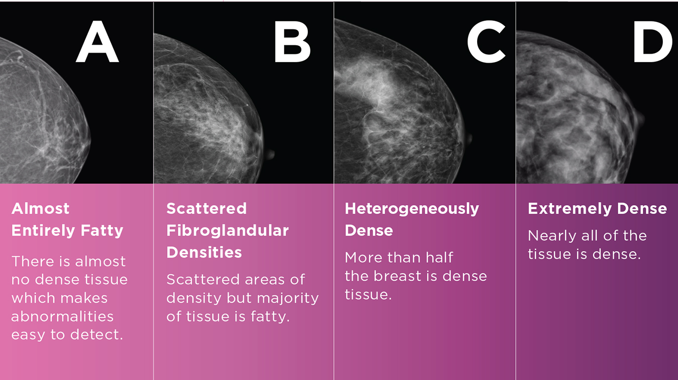
You can’t tell by looking at them, whether or not you have dense breasts. It is a clinical diagnosis that can only be assessed by mammography. Dense breasts have less fat and more glandular and connective tissue. Unfortunately, they also make a mammogram harder to read, so smaller cancers may be hidden. Plus, the denser the breast tissue, the higher the risk of breast cancer.
On a mammogram, fatty tissue looks dark, while both dense tissue and tumours look white, making it hard to distinguish between the two. The white-looking breast cancers are easier to see on a mammogram when they’re surrounded by dark-looking fatty tissue.
Dense breasts are normal. Your breast tissue changes as you age, usually becoming less dense as you get older and go through menopause, but some women continue to have dense breast regardless of age. Dense breasts may be affected by taking hormone replacement therapy.
To determine your breast density, you will need to discuss scheduling a mammogram and a review of the results with your health care provider.
The Breast Imaging Reporting and Database Systems, or BI-RADS, classifies breast density into four groups:
While scoring is not an exact science and radiologists often disagree about levels of density, it is important to get screened. Mayfair uses Volpara software, which scores density from A to D. We will also explain your breast score at the end of your appointment, if requested.
When an assessment determines that breast density is high, the radiologist may suggest annual mammography exams. In addition, the radiologist may suggest the use of handheld or automated breast ultrasound (ABUS) in conjunction with your regular screening mammogram.
Mammography views the breast in slices and provides a greater level of detail, while breast ultrasound increases the sensitivity of the scan. In Alberta, the Toward Optimized Practice Breast Cancer Screening Guidelines suggest that ultrasound “May be used as a supplemental tool by a radiologist after considering current and prior imaging (if available), and history.” You will need to discuss your results and next steps with your health care provider.
Mayfair has 13 mammography clinics in Calgary and one in Cochrane. We also have 13 ultrasound locations in Calgary and one in Cochrane, which offer breast ultrasound services. ABUS is offered at our Market Mall, Mayfair Place, Southcentre, and The CORE locations.
REFERENCES
Alberta Breast Cancer Screening Clinical Practice Guideline Committee (2022) “Alberta Breast Cancer Screening: Clinical Practice Guideline 2022 Update.” www.screeningforlife.ca. Accessed August 12, 2022.
Canadian Cancer Society (2022) “Breast density.” www.cancer.ca. Accessed September 1, 2022.
Canadian Cancer Society (2022) “Risks for breast cancer.” www.cancer.ca. Accessed September 1, 2022.
Łuczyńska, E., et al. (2022) “The Role of ABUS in The Diagnosis of Breast Cancer.” The Journal of Ultrasonography, 2022 Apr; 22(89): 76–85.
Mayo Clinic Staff (2022) “Dense breast tissue: What it means to have dense breasts.” www.mayoclinic.org. Accessed September 1, 2022.
Thigpen, D., et al. (2018) “The Role of Ultrasound in Screening Dense Breasts—A Review of the Literature and Practical Solutions for Implementation.” Diagnostics (Basel), 2018 Mar; 8(1): 20.
We foster a supportive and collaborative culture designed to encourage positive patient experiences and build strong working relationships across the organization:
Our core values shape the way we work with patients, partners, and fellow employees. And, more than anything else, they’re what set Mayfair apart. In everything we do, this is what we strive for:
EXCELLENCE
We share a commitment to high quality and excellence in all that we do. This commitment calls on all of us to achieve the very best of our capabilities and exceed our own expectations.
CURIOSITY
We innovate in everything, from services to processes. We believe meaningful change and effective problem solving come only by looking at challenges and opportunities from new angles and by exercising our creativity and curiosity.
PASSION
We show pride, enthusiasm, and dedication in everything that we do. We are committed to producing and delivering high-quality results and services. We are passionate about our industry and about our company, services, partners, and patients.
COLLABORATION
Our team is supportive of each other’s efforts; we are loyal to one another; and we care for one another both personally and professionally. We promote and support a diverse, yet unified, team. We work together to meet our common goals across Mayfair clinics, locations, and geographies. Only through collaboration on ideas, technologies, and talents can we achieve our mission and vision.
SERVICE
We take pride in delivering exceptional service every day. We listen to every request with an open mind, always looking for opportunities to go above and beyond to create memorable, personalized experiences. We take responsibility to answer our referrers’ and patients’ requests and respect their time by always responding with a sense of urgency.
Start a career with Mayfair Diagnostics — one of Western Canada’s leading medical imaging teams.
Headquartered in Calgary, Alberta, we’ve been helping people f ind clarity for their health for over 100 years. At our clinics in Calgary and area, Regina, and Saskatoon, our team of radiologists, technologists, and support staff work in a truly integrated way to provide exceptional experiences for our patients. Joining our team is more than a job. It’s an investment in your future — a plan for success.
OUR PEOPLE
Our people share our quest to make a difference in our patient’s lives. We’re a team of professionals, disciplined in our skills and compassionate with our patients, providing the care and attention they need. At our core, we are a trusted partner in our patients’ health care journey. Our patients, physicians, and other health care providers rely on us for quality imaging to help manage their patient’s health care decisions with certainty. But our business is about more than just imaging. It’s about building lasting relationships and making a meaningful difference in the lives of those we meet.
OUR VISION
A world in which every person has clarity about their health. We push the boundaries of what is possible and embrace change as an opportunity. We strive to be thought leaders and encourage creativity by providing a safe place for calculated risk taking. We learn from our mistakes. We share best practices across our operations and are recognized by our peers for our work. We engage the best to help propel us forward in achieving our goals.