
Your feet act as shock absorbers that protect the body with every step, accumulating over one million pounds of force, on average, every day. Along with your ankles, they are also incredibly complex mechanisms – that’s why it’s so important to keep them healthy.
Each foot contains 26 bones, 33 joints, and more than a hundred muscles, ligaments, and tendons. The ankle is often referred to as one joint, but it actually accounts for three of those 33 joints. That is a lot of moving parts, some of which are very small.
While the complexity of your feet helps make them strong and agile, it can make treating injuries a challenge. With the amount of stress your feet experience on a daily basis, an accurate diagnosis and appropriate treatment for any concern is paramount. Your health care practitioner will examine the outside of your foot to look for external signs of injury, such as swelling or deformities, but for a specific diagnosis it is usually necessary to take a look inside. A standard X-ray can confirm a bone fracture or arthritis damage, but a more detailed look might require a bone scan.
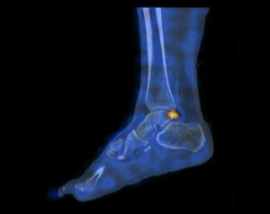
For complex cases, a type of nuclear medicine imaging called a bone scan with SPECT/CT (single photon emission computed tomography/computed tomography) is often recommended. This type of exam can provide detailed pictures of the area of concern and localized information about each specific bone or even specific regions of particular bones. This makes it particularly useful in examining areas with many small bones and joints like the foot and ankle.
A bone scan with SPECT/CT can look at both the structure and function of the area of concern, offering high diagnostic accuracy. For example, when foot and ankle surgery is indicated exact localization of arthritis, stress fractures, and other bone conditions is necessary for optimal results.
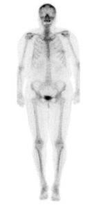
A bone scan with SPECT/CT involves two separate appointments booked on the same day. The first appointment will take approximately 15 minutes. During this time, a small amount of radioactive material is injected; usually into an arm vein, and through blood flow is delivered to the bones in the area of concern. A gamma camera detects the location of the material, taking pictures in a “planar” format.
During the second appointment, imaging will be performed without any additional injections. It will take approximately 30 minutes. A SPECT/CT exam combines a “SPECT” scan with a “CT” scan to help localize the area of abnormal activity that may be present on the planar bone scan image. For the “SPECT” part, the nuclear medicine gamma camera rotates 360 degrees around the body and forms pictures, which the system reconstructs into an image. For the “CT” portion, a low-dose CT image is taken, similar to those from a classic diagnostic CT scan. In this case, they are fused electronically with the SPECT images to get the SPECT/CT image.
This exam is covered under your Alberta Health Care Insurance Plan and must be requested by a health care practitioner. To determine whether it’s appropriate for you, your doctor will often review your medical and family history, risk factors, how long symptoms have been present, and how they affect daily activities. If this exam is indicated as a best next course of action, your doctor will provide you with a requisition and the appointment can be booked.
These exams are performed at our Castleridge, Mahogany Village, Market Mall, Mayfair Place, and Sunpark locations.
It’s important to note that this exam involves a small dose of ionizing radiation from the radiopharmaceutical injected into your vein, and also from the CT scan. CT imaging is a form of X-ray and the exposure to radiation from this scan is slightly higher than that of standard X-rays, but the associated risk is still small. Overall, the radiation exposure from a bone scan with SPECT/CT is about the equivalent of exposure to the earth’s natural background radiation over two years. In most cases, the benefits of a CT, such as the early detection of a serious illness, outweigh the small increased risk from radiation exposure.
Canadian Podiatric Medical Association (2022) “Foot Health.” www.podiatrycanada.org. Accessed October 19, 2022.
Eelsing, R. et al. (2020) “The added value of SPECT/CT in the painful foot and ankle: A review of the literature.” Foot and Ankle Surgery, Vol. 27 (7): 715-722. Accessed October 19, 2022.
Horisberger, M. et al. (2015) “Nuclear Medicine Imaging of Ankle Injuries.” Nuclear Medicine and Radiologic Imaging in Sports Injuries. January 2015: p. 803-816. Accessed October 19, 2022.