Home ![]() IMMIGRATION X-RAY FOR TUBERCULOSIS
IMMIGRATION X-RAY FOR TUBERCULOSIS
Some estimate that tuberculosis (TB) has been around for millions of years, although it’s had many names over the years. In the 1800s, it was known as consumption, and was often still referred to this way despite being formally named tuberculosis in 1834.
TB is an acute or chronic bacterial infection that mostly affects the lungs, but can also affect the brain, kidneys, or spine. It can be caused by two types of bacteria:
When Dr. Robert Koch discovered the bacteria that causes TB in 1882 the disease was killing one out of seven people living in the United States and Europe at that time. Centuries later the disease is still around, and World Tuberculosis Day on March 24 helps educate the public about the impact of TB.
TB bacteria can cause the formation of small tissue masses, called tubercles. When formed in the lungs, these tubercles can impair breathing and lead to coughing and release of sputum. Depending on the area of the body affected and the severity of the infection, symptoms of TB disease can also include feelings of weakness, weight loss, fever, night sweats, chest pain, and the coughing up of blood. If left untreated, TB can lead to death.
Tuberculosis can be inactive (latent) or active:
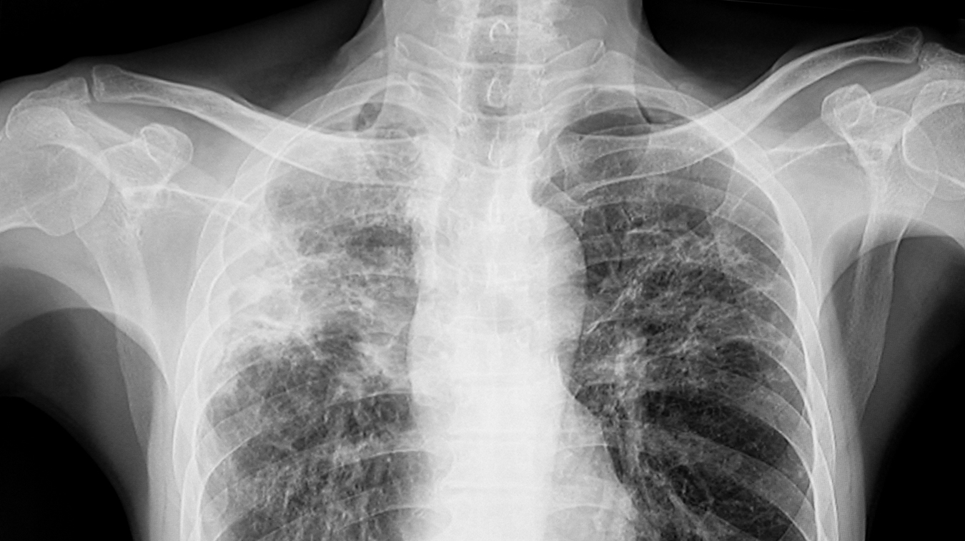
X-ray imaging is used to look at the lungs to check for signs of active TB. X-rays use a type of electromagnetic ionizing radiation to create images of your lungs, heart, and the bones of your chest and spine. A small amount of radiation is sent into the chest and different areas of the chest absorb this radiation at different rates, creating an image with various shades of grey. When filled with air, the lungs show up as darker areas while the heart and lung vessels will appear as lighter areas.
Different lung and heart pathologies can be determined by the radiologist depending on their appearance within the lungs. For example, signs of infection, such as lesions, masses, or fluid, can show up as white patchy areas.
A chest X-ray is a mandatory requirement from Immigration, Refugees, and Citizenship Canada (IRCC). It’s used to check for indications of tuberculosis for anyone looking to immigrate into Canada.
If someone applies to stay in Canada, or if they are emigrating to another country, there are usually a set of admission criteria that include meeting certain medical standards. Within Canada, applicants must not create a danger to public health or public safety.
The chest X-ray will be examined for:
Applicants must bring their immigration medical examination, unique medical identifier, or unique client identifier numbers with them to their X-ray exam. These numbers, or your name and passport numbers, can be used to retrieve your health case in eMedical – an online tool used by doctors, who are approved by IRCC to do medical exams, to record and send immigration medical exam results to IRCC.
In Calgary, immigration chest X-rays for applicants trying to immigrate into Canada can only be requested by IRCC-approved doctors. One of these doctors will give you a requisition form and other paperwork outlining your need for this exam. You will then be directed to go to one of the clinics approved to perform these exams: Mayfair Place, South Calgary Health Centre, The CORE, and Westbrook. These X-rays are offered on a walk-in basis, appointments are not required; simply bring your forms with you.
For applicants trying to immigrate to New Zealand and Australia, these immigration chest X-rays can only be performed at Mayfair Place.
Your X-ray images will be reviewed by a specialized radiologist who will compile a report that is sent to your doctor and uploaded to eMedical. There is a cost for this exam, which can be paid with Visa, MasterCard, debit, or cash.
During this exam, you will be asked to change into a gown and to remove any metal objects, such as jewelry, watches, hair clips, pins, and clothes with metal buttons, zippers, or snaps. We will also ask you to tie up any long hair that may obstruct a clear view of the chest.
For women between the ages of 11-55, you will be asked about the possibility of pregnancy prior to your exam, since X-rays use radiation. Lead shielding can also be provided to cover vulnerable areas. If you are pregnant, you will be asked to sign a consent form, explaining the radiation risks, before proceeding with the exam.
If you require assistance changing or moving, please bring someone to help you, if possible. You will be asked to apply two lead ball nipple markers to distinguish between nipple shadows and lesions in the chest. These are held on with adhesive and are easily removed after the exam.
One of our experienced technologists will help you move into different positions standing with your chest against an upright board. You will be asked to take a deep breath and hold it while the X-ray is being taken. Our state-of-the-art imaging technology will take views of the front and side of your chest. You will need to remain very still while the X-ray is being taken. You will also be required to take a deep breath and hold it for several seconds to help your heart and lungs show up more clearly. We need the lungs filled with air in order to assess them.
The X-ray itself is not painful but holding a particular position may be uncomfortable if you are experiencing pain. Overall, the exam may take about 15 minutes, although once in the X-ray room the process typically takes less than five minutes. Once the technologist has confirmed that the image is of good quality, you will be free to leave. For out-of-town patients, the radiologist will review the images before you leave the clinic.
REFERENCES
Centers for Disease Control and Prevention (2011) “Tuberculosis (TB).” www.cdc.gov. Accessed February 18, 2022.
Centers for Disease Control and Prevention (2016) “History of World TB Day.” www.cdc.gov. Accessed February 18, 2022.
Government of Canada (2020) “Canadian Panel Member Guide to Immigration Medical Examinations 2020.” www.canada.ca. Accessed February 18, 2022.
Healthwise Staff (2020) “Chest X-Ray.” www.myhealth.alberta.ca. Accessed February 18, 2022.
Nabili, S. N. (2020) “What Does a Chest X-Ray Show?” www.medicinenet.com. Accessed February 18, 2022.
National Organization for Rare Disorders (2021) “Tuberculosis.” www.rarediseases.org. Accessed February 18, 2022.
We foster a supportive and collaborative culture designed to encourage positive patient experiences and build strong working relationships across the organization:
Our core values shape the way we work with patients, partners, and fellow employees. And, more than anything else, they’re what set Mayfair apart. In everything we do, this is what we strive for:
EXCELLENCE
We share a commitment to high quality and excellence in all that we do. This commitment calls on all of us to achieve the very best of our capabilities and exceed our own expectations.
CURIOSITY
We innovate in everything, from services to processes. We believe meaningful change and effective problem solving come only by looking at challenges and opportunities from new angles and by exercising our creativity and curiosity.
PASSION
We show pride, enthusiasm, and dedication in everything that we do. We are committed to producing and delivering high-quality results and services. We are passionate about our industry and about our company, services, partners, and patients.
COLLABORATION
Our team is supportive of each other’s efforts; we are loyal to one another; and we care for one another both personally and professionally. We promote and support a diverse, yet unified, team. We work together to meet our common goals across Mayfair clinics, locations, and geographies. Only through collaboration on ideas, technologies, and talents can we achieve our mission and vision.
SERVICE
We take pride in delivering exceptional service every day. We listen to every request with an open mind, always looking for opportunities to go above and beyond to create memorable, personalized experiences. We take responsibility to answer our referrers’ and patients’ requests and respect their time by always responding with a sense of urgency.
Start a career with Mayfair Diagnostics — one of Western Canada’s leading medical imaging teams.
Headquartered in Calgary, Alberta, we’ve been helping people f ind clarity for their health for over 100 years. At our clinics in Calgary and area, Regina, and Saskatoon, our team of radiologists, technologists, and support staff work in a truly integrated way to provide exceptional experiences for our patients. Joining our team is more than a job. It’s an investment in your future — a plan for success.
OUR PEOPLE
Our people share our quest to make a difference in our patient’s lives. We’re a team of professionals, disciplined in our skills and compassionate with our patients, providing the care and attention they need. At our core, we are a trusted partner in our patients’ health care journey. Our patients, physicians, and other health care providers rely on us for quality imaging to help manage their patient’s health care decisions with certainty. But our business is about more than just imaging. It’s about building lasting relationships and making a meaningful difference in the lives of those we meet.
OUR VISION
A world in which every person has clarity about their health. We push the boundaries of what is possible and embrace change as an opportunity. We strive to be thought leaders and encourage creativity by providing a safe place for calculated risk taking. We learn from our mistakes. We share best practices across our operations and are recognized by our peers for our work. We engage the best to help propel us forward in achieving our goals.