
Home ![]() VASCULAR ULTRASOUND FOR POPLITEAL ARTERY ENTRAPMENT
VASCULAR ULTRASOUND FOR POPLITEAL ARTERY ENTRAPMENT
Popliteal artery entrapment syndrome (PAES) is an uncommon vascular condition that occurs when the calf muscles compress the main artery behind the knee (the popliteal artery). This artery is the main source of blood supply to the leg below the knee.
When the popliteal artery is compressed, blood flow is reduced, and you may experience pain and cramping in the back of the lower leg during exercise. The pain usually fades when you stop exercising, but repeated compression and release of the popliteal artery can eventually cause permanent damage to the artery wall.
You can be born with PAES. In this case, the calf muscle or artery are abnormally positioned during fetal development, which causes the popliteal artery to have an irregular path through the calf and be at risk of compression.
You can also develop it later in life. If your calf muscle is in the correct position but is bigger than normal, this can also cause compression of the artery.
Although PAES can affect both men and women, it’s most common in young male athletes 20-40 years of age with no associated vascular risk factors. You may be at higher risk if you are a runner, bicyclist, or athlete who tries to build muscle fast with weight-training routines or high-intensity circuit training.
The main symptom of PAES is pain or cramping in the calf that is brought on during exercise and improves with rest. Other signs and symptoms may include:
If the nearby vein (popliteal vein) also becomes trapped by the calf muscle, you may have:
Please speak to your health care practitioner if you have any type of leg pain, especially if you have calf or foot cramping during an activity that gets better with rest.
PAES can be challenging to diagnose because it’s relatively uncommon and it affects otherwise healthy young adults. The symptoms can also mimic other conditions, such as knee joint pain or other muscle-related problems. In many cases, a diagnosis is made via medical imaging using vascular ultrasound.
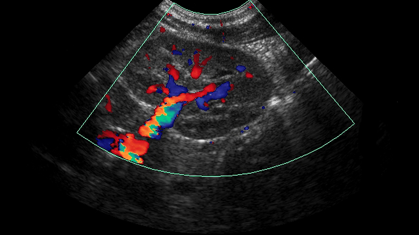
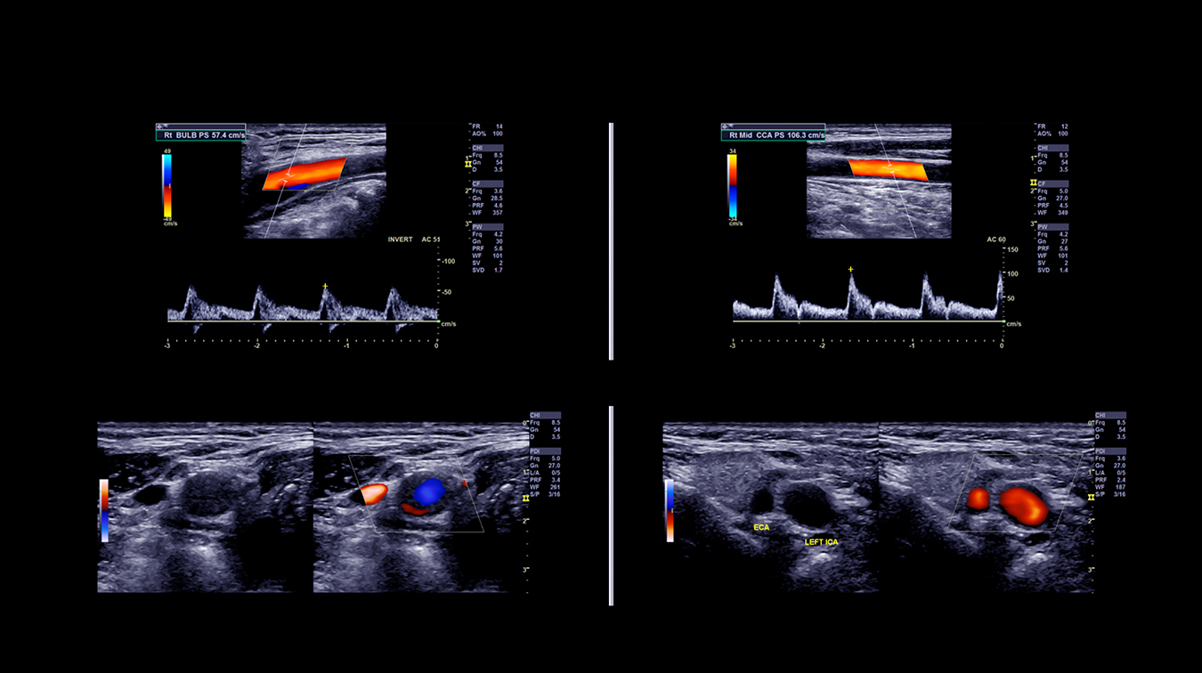
Using sound waves, a vascular ultrasound exam looks at the blood flow in your veins and arteries. High-frequency sound waves are transmitted from a handheld device called a probe or transducer that is placed on the body and moved around the area of interest. A gel is applied to the skin to aid in the movement of the transducer. The transducer collects the sounds that bounce back and a computer then uses those sound waves to create an image on a computer screen. Ultrasound exams are non-invasive, don’t use radiation, and images are captured in real-time, showing the movement of your blood flowing through your blood vessels.
For PAES, a vascular ultrasound exam can assess the popliteal artery with the calf in a neutral or relaxed position and in a flexed position, such as standing on tip toes, toes bent back, or toes pointed forward. Images can then be generated that help determine whether there is any compression or blockage of the popliteal artery that could indicate popliteal artery entrapment syndrome.
This type of vascular ultrasound is safe and usually not painful. Occasionally, it may require pressure to ensure the best image of the area in question and might be temporarily uncomfortable, such as when an area of tenderness is scanned.
After your exam a specialized vascular radiologist will review your images and interpret them before forwarding a report to your referring physician. If there are any concerns with your results, a radiologist may speak directly with you or fax a report to your doctor. For non-urgent results, your doctor will receive a detailed report, which will outline your diagnosis, by the next business day following your exam. Your doctor will then discuss your results with you.
Vascular ultrasound imaging must be requested by a health care practitioner who will provide a requisition. Your medical and family history, risk factors, and type and duration of symptoms, all affect a referring physician’s decision on which type of imaging is appropriate.
When we receive your requisition Mayfair Diagnostics will contact you to schedule your exam and provide you with detailed information to prepare for it. Our more complex vascular studies, such as those to diagnose PAES, are performed at our Vascular Lab at our Sunridge location.
REFERENCES
Hicks, C.W. et al. (2019) “Popliteal Artery Entrapment Syndrome.” Sage Journals. Accessed August 30, 2021.
John Hopkins University (2021) “Popliteal Artery Entrapment Syndrome (PAES).” www.hopkinsmedicine.org. Accessed August 27, 2021.
Mayo Clinic Staff (2021) “Popliteal Artery Entrapment Syndrome.” www.mayoclinic.org. Accessed August 27, 2021.
Radiological Society of North America (2021) “Ultrasound – Vascular.” www.radiologyinfo.org. Accessed August 30, 2021.
We foster a supportive and collaborative culture designed to encourage positive patient experiences and build strong working relationships across the organization:
Our core values shape the way we work with patients, partners, and fellow employees. And, more than anything else, they’re what set Mayfair apart. In everything we do, this is what we strive for:
EXCELLENCE
We share a commitment to high quality and excellence in all that we do. This commitment calls on all of us to achieve the very best of our capabilities and exceed our own expectations.
CURIOSITY
We innovate in everything, from services to processes. We believe meaningful change and effective problem solving come only by looking at challenges and opportunities from new angles and by exercising our creativity and curiosity.
PASSION
We show pride, enthusiasm, and dedication in everything that we do. We are committed to producing and delivering high-quality results and services. We are passionate about our industry and about our company, services, partners, and patients.
COLLABORATION
Our team is supportive of each other’s efforts; we are loyal to one another; and we care for one another both personally and professionally. We promote and support a diverse, yet unified, team. We work together to meet our common goals across Mayfair clinics, locations, and geographies. Only through collaboration on ideas, technologies, and talents can we achieve our mission and vision.
SERVICE
We take pride in delivering exceptional service every day. We listen to every request with an open mind, always looking for opportunities to go above and beyond to create memorable, personalized experiences. We take responsibility to answer our referrers’ and patients’ requests and respect their time by always responding with a sense of urgency.
Start a career with Mayfair Diagnostics — one of Western Canada’s leading medical imaging teams.
Headquartered in Calgary, Alberta, we’ve been helping people f ind clarity for their health for over 100 years. At our clinics in Calgary and area, Regina, and Saskatoon, our team of radiologists, technologists, and support staff work in a truly integrated way to provide exceptional experiences for our patients. Joining our team is more than a job. It’s an investment in your future — a plan for success.
OUR PEOPLE
Our people share our quest to make a difference in our patient’s lives. We’re a team of professionals, disciplined in our skills and compassionate with our patients, providing the care and attention they need. At our core, we are a trusted partner in our patients’ health care journey. Our patients, physicians, and other health care providers rely on us for quality imaging to help manage their patient’s health care decisions with certainty. But our business is about more than just imaging. It’s about building lasting relationships and making a meaningful difference in the lives of those we meet.
OUR VISION
A world in which every person has clarity about their health. We push the boundaries of what is possible and embrace change as an opportunity. We strive to be thought leaders and encourage creativity by providing a safe place for calculated risk taking. We learn from our mistakes. We share best practices across our operations and are recognized by our peers for our work. We engage the best to help propel us forward in achieving our goals.