
Home ![]() WHAT IS MRI ENTEROGRAPHY?
WHAT IS MRI ENTEROGRAPHY?
Magnetic resonance imaging (MRI) is a powerful tool for diagnosis and is often considered the best way to detect diseases in their earliest, most treatable stage. Using a strong magnetic field and radiofrequency waves, this technology can examine the soft tissues of the body along with the bones.
MRI enterography is an imaging test that provides a detailed picture of your small intestine. This scan includes the use of oral and/or injectable contrast dye that’s given to further highlight the small intestine.
The exam uses Mayfair Diagnostics’ state-of-the-art equipment to offer a more comfortable imaging experience. It is often ordered when more detail is needed based on results of other studies or when the cause of symptoms is unclear during a physical exam.
MRI enterography can be used to help diagnose the following:
For MRI enterography, you will be asked avoid food or drink (except water) starting at midnight the night before your exam. You will be asked to arrive 90 minutes before your appointment to allow for enough time to start an intravenous line and for the oral contrast to take effect. You will also be asked to wear comfortable clothes free of metal zippers or buttons, including metal-free underwear. It generally takes between 30-60 minutes to complete this exam.
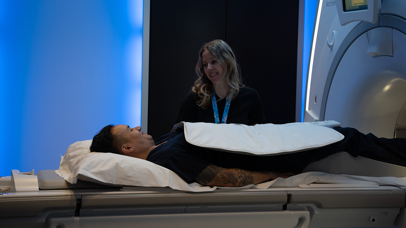
You will be asked to drink roughly one 500 ml bottle of water mixed with 40 ml of Lactulose (an oral contrast solution that helps enhance your images) every 20 minutes over the next 60 minutes. After 60 minutes you will be taken to the MRI scanner and asked to lie on your back on the cushioned scanner table, which will then be adjusted for correct placement. We will do our best to make you comfortable.
An MRI can be loud, so you will be provided with headphones or ear plugs to reduce the noise. Eye masks are available upon request for patients who have difficulty relaxing in a confined space. You are also given an emergency call bell to stop the exam, if needed.
Your technologist will then set up an intravenous (IV) line for the contrast solution. Once you are relaxed and comfortable, a sensor will be positioned around the body part being scanned. The table will then move slowly into the MRI scanner, which is well lit and ventilated.
Your technologist will leave the room to begin your exam, but you will be in direct communication throughout your procedure. Your technologist will take several initial scans, then administer the contrast solution through your IV to enhance your remaining images.
You will be asked to hold very still, as movement may result in blurring of images. Your technologist will be close by at all times. You may be asked to hold your breath for some of the images. Breath holds can last between 5-25 seconds. Once we have successfully captured your images, your IV will be removed and you are free to leave.
An MRI scan creates images by exposing hydrogen atoms in water molecules within our body to a magnetic field which controls the direction and frequency at which hydrogen protons spin. A radio frequency pulse is then directed at a specific area of the body, while smaller magnets are used to alter the magnetic field on a small, but localized level. As tissue responds differently to these magnetic field alterations, a computer can convert the data into a picture.
An MRI’s magnetic field strength is measured in teslas (T). In Alberta, we operate both 1.5T and 3T MRI services at our Mayfair Place location in Calgary. The highly sensitive images from the 3T machine allow enhanced imaging of many areas of the body, including soft tissue.
In Saskatchewan, we operate 1.5T MRI services at our Regina and Saskatoon clinics.
MRI images are created using a magnetic field, which can attract metal objects or may cause metal in your body to move. This means that before an MRI can be performed all patients will need to be screened to exclude internal metal objects or hardware that are not safe in the MRI. The inside of the MRI scanner can feel small to some people and there are noises caused by changes in the magnetic field, which require ear protection.
When using MRI contrast, an injectable gadolinium dye, there is a small risk of allergic reaction. However, you will be asked about your allergies and other medical conditions when booking your exam.
In Alberta, MRI exams are available in hospitals and covered under the Alberta Health Care Insurance Plan, but we also offer the same studies as private pay exams, for when patients may be unable to wait to receive an exam through the public health system. In Saskatchewan, we offer MRIs as a publicly funded, community-based service under contract with the Saskatchewan Health Authority and as a private pay exam. Private MRI services in Saskatchewan are provided in accordance with and under the legislation of the Province of Saskatchewan.
Whether public or private, an MRI must be requested by a health care practitioner who will provide a requisition. Mayfair Diagnostics will schedule your exam and provide you with detailed information to prepare for it. Once your exam is completed, your images will be reviewed by a specialized radiologist who will compile a report that is sent to your doctor.
Mayfair Diagnostics is owned and operated by over 50 radiologists who are sub-specialty trained, which guarantees an expert opinion of your imaging.
Mayfair Diagnostics offers MRI imaging as a private pay service at our Mayfair Place location in Calgary, and as both public and private pay exams at our Regina and Saskatoon locations in Saskatchewan. For more information, please visit our services page or call our toll-free number 1-866-611-2665.
REFERENCES
Healthwise Staff (2022) “Magnetic Resonance (MR) Enterogram: About This Test.” myhealth.alberta.ca. Accessed February 22, 2024.
Johns Hopkins Medicine (2024) “MR Enterography.” www.hopkinsmedicine.org. Accessed February 22, 2024.
Radiological Society of North America (2023) “MR Enterography.” www.radiologyinfo.org. Accessed February 22, 2024.
Saskatchewan Health Authority (2024) “Magnetic Resonance Imaging (MRI).” www.saskatchewan.ca. Accessed February 22, 2024.
Sinha, R., et al. (2011) “MR Enterography of Crohn Disease: Part 1, Rationale, Technique, and Pitfalls.” American Journal of Radiology. June 2011; 197:76-79. Accessed February 22, 2024.
We foster a supportive and collaborative culture designed to encourage positive patient experiences and build strong working relationships across the organization:
Our core values shape the way we work with patients, partners, and fellow employees. And, more than anything else, they’re what set Mayfair apart. In everything we do, this is what we strive for:
EXCELLENCE
We share a commitment to high quality and excellence in all that we do. This commitment calls on all of us to achieve the very best of our capabilities and exceed our own expectations.
CURIOSITY
We innovate in everything, from services to processes. We believe meaningful change and effective problem solving come only by looking at challenges and opportunities from new angles and by exercising our creativity and curiosity.
PASSION
We show pride, enthusiasm, and dedication in everything that we do. We are committed to producing and delivering high-quality results and services. We are passionate about our industry and about our company, services, partners, and patients.
COLLABORATION
Our team is supportive of each other’s efforts; we are loyal to one another; and we care for one another both personally and professionally. We promote and support a diverse, yet unified, team. We work together to meet our common goals across Mayfair clinics, locations, and geographies. Only through collaboration on ideas, technologies, and talents can we achieve our mission and vision.
SERVICE
We take pride in delivering exceptional service every day. We listen to every request with an open mind, always looking for opportunities to go above and beyond to create memorable, personalized experiences. We take responsibility to answer our referrers’ and patients’ requests and respect their time by always responding with a sense of urgency.
Start a career with Mayfair Diagnostics — one of Western Canada’s leading medical imaging teams.
Headquartered in Calgary, Alberta, we’ve been helping people f ind clarity for their health for over 100 years. At our clinics in Calgary and area, Regina, and Saskatoon, our team of radiologists, technologists, and support staff work in a truly integrated way to provide exceptional experiences for our patients. Joining our team is more than a job. It’s an investment in your future — a plan for success.
OUR PEOPLE
Our people share our quest to make a difference in our patient’s lives. We’re a team of professionals, disciplined in our skills and compassionate with our patients, providing the care and attention they need. At our core, we are a trusted partner in our patients’ health care journey. Our patients, physicians, and other health care providers rely on us for quality imaging to help manage their patient’s health care decisions with certainty. But our business is about more than just imaging. It’s about building lasting relationships and making a meaningful difference in the lives of those we meet.
OUR VISION
A world in which every person has clarity about their health. We push the boundaries of what is possible and embrace change as an opportunity. We strive to be thought leaders and encourage creativity by providing a safe place for calculated risk taking. We learn from our mistakes. We share best practices across our operations and are recognized by our peers for our work. We engage the best to help propel us forward in achieving our goals.