TOLL FREE:
1-866-611-2665

Home WHAT’S THE DIFFERENCE BETWEEN CT AND PET IMAGING?
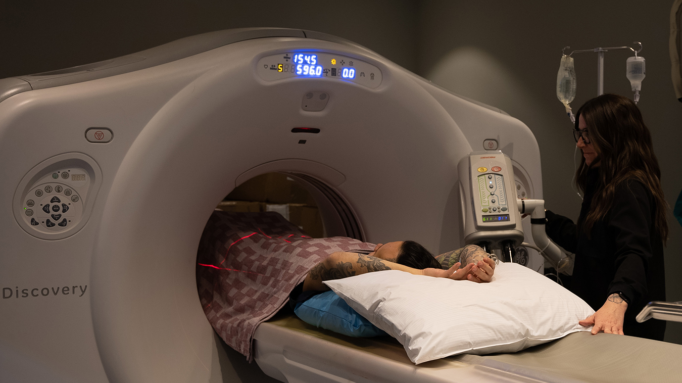
Computed tomography (CT) and positron emission tomography (PET) are medical imaging exams that allow your doctor to get a very detailed look inside your body. They are similar in the way they are performed – you lie on a moving table that passes through a doughnut shaped machine. However, their technology and the information they provide to medical professionals is quite distinct.
CT scans are useful to look at anatomical structures such as organs, bones, and tissues, whereas PET scans measure vital functions such as blood flow, oxygen use, and blood sugar metabolism to show how the tissues work on a cellular level.
There are many uses for a CT scan, but it’s particularly well-suited for evaluating injuries and diagnosing diseases, such as lung or colon cancer and coronary artery disease. Unlike other forms of imaging, a PET scan shows molecular activity to help identify changes occurring in an organ or tissue. They can be used to evaluate the presence of disease or other conditions and assess the function of the brain or heart. The most common use of PET is in the detection of cancer and the evaluation of cancer treatment.
CT scans use a combination of X-rays and computer technology to produce comprehensive images of any part of the body. They are much more detailed than regular X-rays. A standard X-ray machine emits a small amount of radiation photons through the body part of interest. A CT scanner emits a series of beams as it moves 360 degrees around the body. A specialized computer reconstructs the signal into 3D images that can be viewed from multiple directions.
Depending on the area of body being scanned, you may be given oral or intravenous (IV) contrast dye to highlight the area being imaged. Dense areas of the body, such as bones, often show clearly on CT, but soft tissues may require the use of contrast. Contrast can limit X-rays in certain areas, appearing white in the scan and helping to highlight blood vessels, organs, or other structures.
PET scans are a type of nuclear medicine imaging. They use a small amount of a radiopharmaceutical, known as a radiotracer, that is injected intravenously. The radiotracer accumulates in organs and tissues where it gives off gamma rays.
A PET scanner uses a specialized camera that detects the gamma rays emitted from your body and, with the help of a computer, creates detailed images. Areas of the body that have a higher uptake of the radiotracer show up as different degrees of brightness in the scan (“hot” spots), which give information about organ and tissue function. The “hot” spots help to determine where in the body changes are taking place.
CT scans can provide detailed information about soft tissues in the chest or abdomen, and blood vessels. They can evaluate fractures or look for blood clots.
CT lung screening can help examine suspicious lung nodules for lung cancer, as well as other serious illnesses. It could be appropriate for patients at high risk of lung cancer due to smoking, a family or personal history of lung cancer, or other risk factors. In these cases, CT screening can help detect early signs of lung cancer, as small as just a few millimeters in size.
For those at risk of coronary artery disease, a coronary CT angiography can be used to non-invasively examine the coronary arteries. It can detect both calcified or hard plaques and noncalcified or soft plaques. It is these soft plaque deposits, often invisible with standard imaging tools, that are more likely to cause heart attack related health issues.
CT virtual colonoscopy is a minimally invasive CT scan that uses low-dose X-rays to produce two- and three-dimensional images of the large intestine (colon) and rectum, which can be used to screen for colon cancer. It differs from a colonoscopy in that an endoscope is not used and your rectum is inflated with CO2, which is more comfortable and more readily absorbed by the body than room air.
PET scans are valuable for detecting cancer and monitoring its treatment, as well as assessing certain heart and brain issues.
Cancer cells have a higher metabolic rate than noncancerous cells causing them to uptake more of the radiotracer and show up as bright coloured spots on PET scans. This helps when detecting cancer, checking if cancer has spread, if a treatment is working, or for a recurrence of cancer.
For heart problems, PET scans can reveal decreased blood flow in the heart. Healthy heart tissue takes up more radiotracer than the unhealthy tissue or tissue with a decreased blood flood. The different colours and degrees of brightness on the scan indicate the different levels of tissue function.
In brain disorders, glucose is the main fuel of the brain. A PET scan uses a glucose radiopharmaceutical, which helps to detect which areas of the brain are utilizing the glucose at the highest rates. This helps to evaluate how the brain is working and to check for any abnormalities.
Mayfair Diagnostics does not offer PET scans – they are available in Calgary through Alberta Health Services (AHS). You would need to discuss with your doctor about the appropriateness of this exam for your concern, and, if needed, you doctor will provide you with a referral to an AHS facility.
CT scans are also available through the public health care system. Mayfair Diagnostics offers community-based private CT services as a complement to the public health care system. They are offered as a private pay exam at our Mayfair Place location and are not covered by Alberta Health Care.
CT scans can be purchased for single or multiple body areas. We also offer Health Assessment packages, which provide a discount on multiple imaging exams when purchased together.
Your health spending account or group medical insurance plan may cover the cost of a private CT that is prescribed by a qualified health care practitioner. You will need to check with your plan administrator for coverage details.
Whether public or private, a CT must be requested by a health care practitioner. To determine whether a CT is recommended, you will need to discuss with your doctor your medical and family history, risk factors, and if there are symptoms, how long symptoms have been present and how they affect daily activities.
If a private CT scan is indicated as a best next course of action, a requisition will be provided, and the appointment can be booked. It’s important to note that the exposure to radiation from a CT scan is higher than that of standard X-rays, but the associated risk is still small. For example, the radiation exposure from one low-dose CT scan of the chest is less than the exposure from the earth’s natural background radiation over six months. In most cases, the benefits of a CT, such as the early detection of a serious illness, outweigh the small increased risk from radiation exposure.
For more information, please visit our services page.
REFERENCES
Begum, J., & Bhatt Patel, R. (2023) “What is a CT Scan?” webmd.com. Accessed April 22, 2024.
National Institute of Biochemical Imaging and Bioengineering (2022) “Computed Tomography (CT).” nibib.nih.gov. Accessed April 22, 2024.
Neurologica (2021) “PET Scan vs. CT Scan.” neurologica.com. Accessed April 22, 2024.
Radiological Society of North America (2023) “PET/CT.” radiologyinfo.org. Accessed April 22, 2024.
Our Refresh newsletter delivers the latest medical news, expert insights, and practical tips straight to your inbox, empowering you with knowledge to enhance patient care and stay informed.
By subscribing to our newsletter you understand and accept that we may share your information with vendors or other third parties who perform services on our behalf. The personal information collected may be stored, processed, and transferred to a country or region outside of Quebec.
Please read our privacy policy for more details.