
Home ![]() NUCLEAR MEDICINE IMAGING FOR FOOT & ANKLE FRACTURES
NUCLEAR MEDICINE IMAGING FOR FOOT & ANKLE FRACTURES
Your feet and ankles are two of the most complex and hardworking regions of the body. They are stable enough to bear your body weight and resist external forces, as well as working like a spring to push the body up and forward during walking. At the same time, they must be flexible enough to accommodate the different surfaces we walk upon.
While the complexity of your feet helps make them strong and agile, it can make treating injuries a challenge. Your feet act as shock absorbers that protect the body with every step, accumulating over one million pounds of force on average every day.
Each foot contains 26 bones, 33 joints, and more than a hundred muscles, ligaments, and tendons. The ankle is often referred to as one joint, but it actually accounts for three of those 33 joints. That is a lot of moving parts, some of which are very small.
With the amount of stress your feet experience on a daily basis, it’s very common to experience pain in and around your feet or ankles. Most of the time this pain is short term and caused by soft tissue injuries, such as sprains or strains. Other types of pain can be more serious, such as:
These types of pain could be caused by fractures of the foot or ankle. However, the complexity of this area means that while an accurate diagnosis and appropriate treatment for any concern is important, it can also be difficult.
Foot fractures are fairly common and may be caused by falls, twisting, or direct impact of your foot against a hard object. They can cause pain which is made worse by trying to walk or put weight on the foot. Treatment can depend on which bone is fractured and the type of fracture.
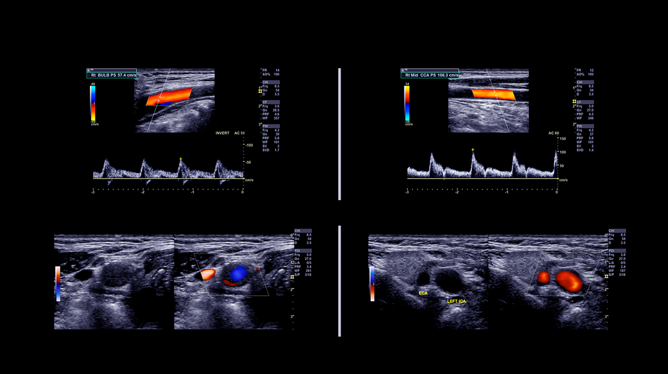
Your health care practitioner will examine the outside of your foot to look for external signs of injury, such as swelling or deformities, but for a specific diagnosis it is usually necessary to take a look inside. A standard X-ray can confirm a bone fracture or arthritis damage, but a more detailed look might require a bone scan.
For complex cases, a type of nuclear medicine imaging called a bone scan with SPECT/CT (single photon emission computed tomography/computed tomography) can be recommended. This type of exam can provide detailed pictures of the area of concern and localized information about each specific bone or even specific regions of particular bones. This makes it particularly useful in examining areas with many small bones and joints like the foot and ankle.
A bone scan with SPECT/CT can look at both the structure and function of the area of concern, offering high diagnostic accuracy. For example, when foot and ankle surgery is indicated exact localization of arthritis, stress fractures, and other bone conditions is necessary for optimal results.
A bone scan with SPECT/CT is a two-part appointment. Part one is usually booked in the morning and will take approximately 15 minutes. During this time, the radiopharmaceutical is injected into an arm vein and travels throughout the body. Through blood flow, it’s delivered to the bones in the area of concern and a gamma camera detects the location of the material, taking pictures in a “planar” format.
You will be asked to lie on your back on the imaging bed while the gamma camera is placed over your body. The first set of images documents increased or decreased flow to the areas of concern. Afterwards, you will be able to go about your normal daily activities until the second part of the appointment (booked 2-4 hours after the first).
During the second part of the appointment, imaging will be performed without any additional injections, to document uptake in the bones. It will take approximately 30-45 minutes. You will be asked to hold as still as possible, while breathing normally – movement can blur the images and make them more difficult to interpret. The SPECT/CT imaging will be performed towards the end of this second appointment.
SPECT/CT (Single Photon Emission Computed Tomography/Computed Tomography) imaging combines two imaging types to help localize the area of abnormal activity that may be present on the planar bone scan image. For the “SPECT” part, the nuclear medicine gamma camera rotates 360 degrees around the body and acquires measurements of the radiation being emitted, which the system reconstructs into a 3D image. For the “CT” portion, a low-dose CT image is taken, similar to those from a classic diagnostic CT scan, but using limited radiation. In this case, they are fused electronically with the SPECT images to get the SPECT/CT image.
This exam is covered under your Alberta Health Care Insurance Plan and must be requested by a health care practitioner. To determine whether it’s appropriate for you, your doctor will often review your medical and family history, risk factors, how long symptoms have been present, and how they affect daily activities. If this exam is indicated as a best next course of action, your doctor will provide you with a requisition and the appointment can be booked.
These exams are performed at our Castleridge, Mahogany Village, Market Mall, Mayfair Place, and Sunpark locations.
It’s important to note that this exam involves a small dose of ionizing radiation from the radiopharmaceutical injected into your vein, and also from the CT scan. CT imaging is a form of X-ray and the exposure to radiation from this scan is slightly higher than that of standard X-rays, but the associated risk is still small. In most cases, the benefits of a bone scan with SPECT/CT, such as the early detection of a serious illness, outweigh the small increased risk from radiation exposure.
Angin, S. & Demirbüken, I. (2020) “Ankle and foot complex.” Comparative Kinesiology of the Human Body, chapter 23, Pages 411-439. Accessed October 25, 2022.
Canadian Podiatric Medical Association (2022) “Foot Health.” www.podiatrycanada.org. Accessed October 25, 2022.
Classen, L. et al. (2014) “Influence on Therapeutic Decision Making of SPECT-CT for Different Regions of the Foot and Ankle.” BioMed Research International, Volume 2014. Accessed October 25, 2022.
Horisberger, M. et al. (2015) “Nuclear Medicine Imaging of Ankle Injuries.” Nuclear Medicine and Radiologic Imaging in Sports Injuries. January 2015: p. 803-816. Accessed October 25, 2022.
Singh, V.K., et al. (2013) “The diagnostic value of single photon-emission computed tomography bone scans combined with CT (SPECT-CT) in diseases of the foot and ankle.” Foot and Ankle Surgery, vol. 19 (2): 80-83. Accessed October 25, 2022.
Versus Arthritis (2022) “Why do I have pain in my foot or ankle?” www.versusarthritis.org. Accessed October 25, 2022.
We foster a supportive and collaborative culture designed to encourage positive patient experiences and build strong working relationships across the organization:
Our core values shape the way we work with patients, partners, and fellow employees. And, more than anything else, they’re what set Mayfair apart. In everything we do, this is what we strive for:
EXCELLENCE
We share a commitment to high quality and excellence in all that we do. This commitment calls on all of us to achieve the very best of our capabilities and exceed our own expectations.
CURIOSITY
We innovate in everything, from services to processes. We believe meaningful change and effective problem solving come only by looking at challenges and opportunities from new angles and by exercising our creativity and curiosity.
PASSION
We show pride, enthusiasm, and dedication in everything that we do. We are committed to producing and delivering high-quality results and services. We are passionate about our industry and about our company, services, partners, and patients.
COLLABORATION
Our team is supportive of each other’s efforts; we are loyal to one another; and we care for one another both personally and professionally. We promote and support a diverse, yet unified, team. We work together to meet our common goals across Mayfair clinics, locations, and geographies. Only through collaboration on ideas, technologies, and talents can we achieve our mission and vision.
SERVICE
We take pride in delivering exceptional service every day. We listen to every request with an open mind, always looking for opportunities to go above and beyond to create memorable, personalized experiences. We take responsibility to answer our referrers’ and patients’ requests and respect their time by always responding with a sense of urgency.
Start a career with Mayfair Diagnostics — one of Western Canada’s leading medical imaging teams.
Headquartered in Calgary, Alberta, we’ve been helping people f ind clarity for their health for over 100 years. At our clinics in Calgary and area, Regina, and Saskatoon, our team of radiologists, technologists, and support staff work in a truly integrated way to provide exceptional experiences for our patients. Joining our team is more than a job. It’s an investment in your future — a plan for success.
OUR PEOPLE
Our people share our quest to make a difference in our patient’s lives. We’re a team of professionals, disciplined in our skills and compassionate with our patients, providing the care and attention they need. At our core, we are a trusted partner in our patients’ health care journey. Our patients, physicians, and other health care providers rely on us for quality imaging to help manage their patient’s health care decisions with certainty. But our business is about more than just imaging. It’s about building lasting relationships and making a meaningful difference in the lives of those we meet.
OUR VISION
A world in which every person has clarity about their health. We push the boundaries of what is possible and embrace change as an opportunity. We strive to be thought leaders and encourage creativity by providing a safe place for calculated risk taking. We learn from our mistakes. We share best practices across our operations and are recognized by our peers for our work. We engage the best to help propel us forward in achieving our goals.