

Home  CYST VS. TUMOUR: WHAT’S THE DIFFERENCE?
If you find a lump or bump, it may be a cyst or tumour. These are two common types of lumps which look similar, but have distinct characteristics.
Cysts can appear anywhere in the body, but most frequently occur in the skin, ovaries, breasts, or kidneys. They are fluid-filled sacs that are usually benign (noncancerous). They can vary in size, from as small as a grain of rice to as large as a golf ball, and there may be more than one cyst present. They can form for no apparent reason but may be caused by blocked ducts or an injury.
Breast cysts typically occur due to hormonal fluctuations during a woman’s menstrual cycle, making them most common in women between the ages of 30 and 50. Breast cysts do not increase the risk of developing breast cancer and often do not require treatment.
Tumours form when a mass of abnormal cells accumulates in tissue. Unlike cysts, tumours are solid masses and can either be benign (noncancerous) or malignant (cancerous).
Differences between cysts and tumours can be difficult to determine without medical attention. It’s important to consult with your doctor about any changes in your body, including new lumps and pain.
To diagnose lumps and cysts, your doctor will likely first perform a physical exam and then order medical imaging, such as ultrasound, mammography (for lumps and cysts in breast tissue), or both together.
An ultrasound uses high-frequency sound waves to help determine the composition of a lump or area of concern, distinguishing between a cyst, fibroglandular tissue, and a solid mass. The pitch, direction, and distance sound waves travel differ depending on what they run into (e.g., tissue, fluid, bone). A computer can interpret this information as a two-dimensional image on a screen and provide information about the area of concern.
For example, an ultrasound can help determine whether the cyst is filled with fluid, solid areas, or a combination of both. By examining the features of the cyst as presented on the ultrasound image, the radiologist can assess whether the cyst has features that may be concerning and might require a biopsy.
Mammography uses X-ray technology to create a detailed picture of the internal structure of breast tissue. This exam can help spot a breast cyst, calcifications, masses, changes in the breast tissue or an area that needs further investigation. When investigating lumps or cysts, a breast ultrasound may also be ordered to complement the mammogram and allow for more comprehensive imaging of the area in question.
A biopsy is a procedure that removes small pieces of tissue from within the breast. A needle is guided into the area of concern to take a small tissue sample, which is sent to a laboratory for analysis.
During mammography, the machine will gently press down on the breasts to spread the breast tissue out and capture a more complete picture of each breast. The pressure lasts for a few seconds, while the machine quickly takes a number of pictures. Then the process will be repeated for the other breast. It may be a bit uncomfortable, but it’s very quick, only 10 or 15 minutes in total.
All Mayfair Diagnostics’ mammography clinics use technology that provides 3D images (tomosynthesis) of the breast that can then be viewed in slices. This provides a greater level of detail and a clearer view of the breast tissue with a very small dose of radiation.
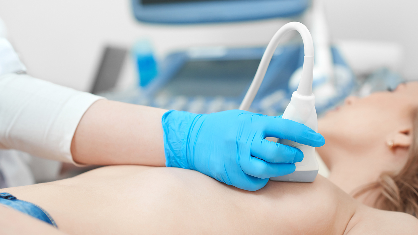
During an ultrasound, a warm, non-scented, hypo-allergenic ultrasound gel will be applied to the area in question. The sonographer moves the handheld probe slowly over the area. In the case of breast ultrasound, this will likely include all areas of each breast and into the armpit area to provide a complete set of images. A general ultrasound takes about 20 minutes, while breast ultrasound can take up to an hour to scan both breasts.
For both exams, you may want to let your technologist know the location of any lumps or areas of pain.
Once the mammogram and/or ultrasound images have been taken, one of our radiologists will look over them very carefully to check for possible abnormalities or changes compared to previous ultrasounds or mammograms.
If you have pain or a concern about a lump, you will need to speak to your doctor about your family history, your medical history, and whether ultrasound, mammography, or both are needed. Your doctor will fill out a requisition for the appropriate imaging and fax it to us, or you can call to book the appointment yourself.
Breast cysts don’t increase your risk of breast cancer, but they may complicate breast screening if you have them or develop them regularly. On a mammogram, breasts cysts may make it challenging to monitor or find changes in breast tissue. For this reason, it’s recommended that you:
If you have breast cysts or develop them regularly, your doctor may recommend yearly breast screening that includes both mammography and breast ultrasound. Regular breast cancer screening through mammography is considered the best way to assess the health of breast tissue and catch breast cancer early in its early, most treatable stage.
Many people start having regular mammograms every year at age 40, since Alberta Health Care covers one mammogram per year starting at that age. The Canadian Association of Radiologists and Mayfair Diagnostics recommend screening mammography every year from age 40 to 49, then every two years between age 50 and 75, if there are no risks factors that would necessitate a shorter interval. After age 75, screening frequency will depend on many factors, including your medical history.
Refer to the Alberta and Saskatchewan provincial breast screening programs for their screening recommendations.
In Alberta, if your doctor hasn’t told you that you need a mammogram, you can still book your exam. You don’t always need a doctor’s requisition to book a screening mammogram. In certain instances, you are able to self-refer. See mammogram self-referral guidelines.
Mayfair Diagnostics has 13 ultrasound and mammography locations in Calgary, one in Cochrane, and one in Regina. Our Cochrane location and 12 of our Calgary locations use the Senographe Pristina mammography system – which helps provide a more comfortable mammogram. Visit our breast imaging page for more information.
REFERENCES
Canadian Cancer Society (2024) “What is breast cancer?” cancer.ca. Accessed July 11, 2024.
Canadian Cancer Society (2024) “Breast cysts.” cancer.ca. Accessed July 10, 2024
Demarco, C. (2022) “Breast cysts and breast cancer: How can you tell the difference?” mdanderson.org. Accessed July 10, 2024.
Radiological Society of North America (2024) “Breast Cancer – Diagnosis, Evaluation and Treatment” radiologyinfo.org. Accessed July 15, 2024.
Smith, C. (2020) “Tumour vs Cyst: Differences, Diagnosis and Treatments.” treatcancer.com. Accessed July 19, 2024.
Victoria State Government, Department of Health (2014) “Cysts.” betterhealth.vic.gov.au. Accessed July 16, 2024.
Radiologists are specialized physicians who interpret diagnostic imaging to diagnose and monitor a wide range of medical conditions. At Mayfair Diagnostics, they review X-ray, ultrasound, CT, and MRI studies, among others, analyzinsg images in detail and providing comprehensive reports and clinical recommendations to referring physicians. They collaborate closely with technologists and clinic teams to guide imaging protocols, ensure quality and radiation safety standards, and may perform image-guided procedures such as biopsies or injections. Through their expertise and teamwork, radiologists play a key role in accurate diagnosis, effective treatment planning, and improved patient outcomes.
Administrative professionals support the organization across human resources, marketing, operations, strategic partnerships, finance, information technology, and infrastructure. From recruiting staff to promoting services and improving workflows, they help ensure smooth operations and positive experiences for employees and patients. Through collaboration with clinical and support teams, they provide essential coordination that enables efficient, high-quality service.
Diagnostic Imaging Assistant (DIA) support clinic operations and help ensure a positive patient experience. They assist staff by greeting and preparing patients, confirming information, coordinating appointments, and guiding patients through their visit. DIAs also maintain exam rooms, manage documentation, and ensure supplies and equipment are ready. Through strong customer service, attention to detail, and teamwork, they help create a safe and organized environment.
Patient Experience Coordinators (PECs) are the first point of contact, scheduling exams and ensuring accurate patient information. They communicate clearly with patients, coordinate with care teams, and support a smooth, confidential, and customer-focused experience.
Nuclear Medical Technologists perform diagnostic nuclear medicine procedures involving sensitive and highly personal patient circumstances. They are responsible for delivering the highest standard of care in a professional, compassionate, and patient-centered manner, in accordance with provincial regulatory requirements, CAMRT standards, and Mayfair policies and guidelines.
Computed Tomography (CT) Technologists operate CT imaging equipment to produce detailed cross-sectional images of the body that assist in diagnosing a wide range of medical conditions. They prepare and position patients for scans, ensure safety protocols are followed, and administer contrast agents when required. CT technologists work closely with radiologists to ensure high-quality diagnostic images are obtained, while providing clear communication and compassionate care to support patient comfort throughout the procedure.
Magnetic Resonance Imaging (MRI) Technologists operates MRI scanners to produce detailed images of internal body structures used to assist in medical diagnosis and treatment planning. They are responsible for preparing and positioning patients, ensuring all safety protocols are strictly followed due to the strong magnetic field, and obtaining high-quality images as directed by radiologists. MRI technologists combine technical expertise with patient care, providing clear communication and support to ensure a safe, comfortable, and efficient imaging experience.
Diagnostic Medical Sonographers perform ultrasound exams to help diagnose and monitor medical conditions. Following Sonography Canada standards and Mayfair Diagnostics protocols, they capture accurate images while ensuring patient safety, comfort, and confidentiality. They work with radiologists and clinical teams to review requisitions, prepare patients, perform scans, and document findings, contributing to accurate diagnoses and a positive patient experience.
Medical Radiation Technologists (MRTs) perform x-ray, mammography, and BMD exams while ensuring patient safety, accuracy, and compassionate care. They also may assist in pain therapy procedures. MRTs verify patient information, explain procedures, position patients, and produce high-quality images. MRTs follow professional standards and protocols, maintaining strict radiation safety, quality assurance, and patient privacy while supporting a positive patient experience.
We foster a supportive and collaborative culture designed to encourage positive patient experiences and build strong working relationships across the organization:
Our core values shape the way we work with patients, partners, and fellow employees. And, more than anything else, they’re what set Mayfair apart. In everything we do, this is what we strive for:
EXCELLENCE
We share a commitment to high quality and excellence in all that we do. This commitment calls on all of us to achieve the very best of our capabilities and exceed our own expectations.
CURIOSITY
We innovate in everything, from services to processes. We believe meaningful change and effective problem solving come only by looking at challenges and opportunities from new angles and by exercising our creativity and curiosity.
PASSION
We show pride, enthusiasm, and dedication in everything that we do. We are committed to producing and delivering high-quality results and services. We are passionate about our industry and about our company, services, partners, and patients.
COLLABORATION
Our team is supportive of each other’s efforts; we are loyal to one another; and we care for one another both personally and professionally. We promote and support a diverse, yet unified, team. We work together to meet our common goals across Mayfair clinics, locations, and geographies. Only through collaboration on ideas, technologies, and talents can we achieve our mission and vision.
SERVICE
We take pride in delivering exceptional service every day. We listen to every request with an open mind, always looking for opportunities to go above and beyond to create memorable, personalized experiences. We take responsibility to answer our referrers’ and patients’ requests and respect their time by always responding with a sense of urgency.
Join Mayfair Diagnostics, recognized as one of Western Canada’s premier medical imaging organizations. With a century-long legacy and headquartered in Calgary, Alberta, Mayfair Diagnostics is dedicated to assisting patients in achieving clarity regarding their health.
Operating clinics in Calgary and surrounding areas, Regina, and Saskatoon, our multidisciplinary team of radiologists, technologists, and support staff collaborate seamlessly to deliver high quality patient care. Working here is more than just a job; it’s a strong step towards your future.
OUR TEAMS
We are a dedicated group of professionals who combine skill with compassion to deliver attentive care to our patients. As a reliable partner in their health care journey, we provide high-quality imaging that assists patients, physicians, and other providers in making informed health decisions. Our work goes beyond imaging; it’s about fostering relationships and positively impacting everyone we serve.
OUR VISION
We envision a world where every person has clarity about their health. We innovate and welcome change, promoting leadership and creativity through safe risk-taking. We share best practices, earn peer recognition for our work, and engage top talent to reach our goals.
We strive to be thought leaders and encourage creativity by providing a safe place for calculated risk taking.
We share best practices across our operations and are recognized by our peers for our work. We engage the best to help propel us forward in achieving our goals.